Today I am presenting routine radiographs of a 77-year-old man with previous history of carcinoma of the bladder ten years ago. What do you see?
Check the images below, leave your thoughts in the comments section, and come back on Friday for the answer.
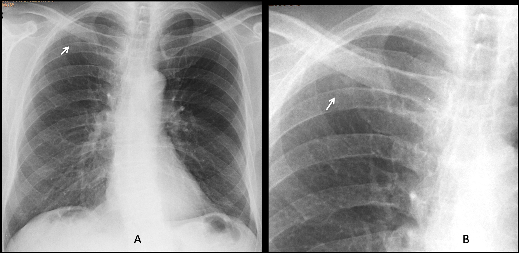
Findings: the PA chest radiograph shows a small right infraclavicular nodule (A-B, arrows), that was overlooked.
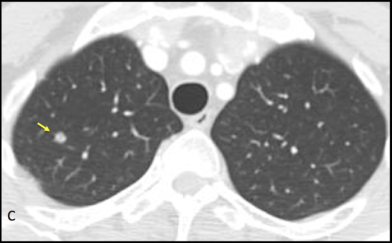
Interestingly enough, a CT taken six months earlier shows the nodule, smaller in size, that was also overlooked (C, arrow).
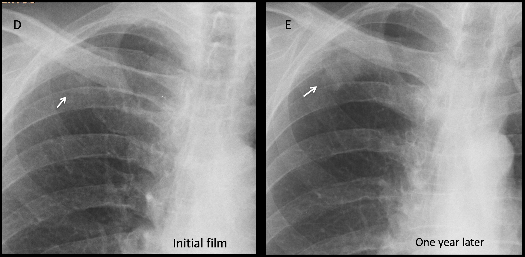
One year later the nodule has increased markedly in size (D-E, arrows) and this time it was noticed.
Considering the clinical history, the most likely diagnosis is carcinoma of the lung, which is frequently associated to carcinoma of the bladder. A metastasis is less likely, given the time interval since the original bladder surgery. The patient was operated on and a malignant melanoma was found.
Final diagnosis: melanoma of the lung.
Congratulations to Katarzyna who was the first one to see the nodule.
Teaching point: up to 50% of apical pulmonary nodules are missed in the chest radiograph. Remember to look carefully at the apices in every examination because many of them are malignant.


Good morning, professor!! We see an hipertransparency in left lung with reduction of the hilar vessels. Tumoral embolism? Does the patient have any respiratory symptoms?
Greetings
Lucretius
This was a routine examination. The patient was asymptomatic. Not only the left lung but the whole left hemithorax and shoulder are blacker than the opposite one. This is due to technical factors and it is a common cause of apparent hyperlucent lung.
Hello dear professor,
finding are following:
– gas under right diaphragm – seems as colonic interposition as in Chilaiditi syndrome – lack of symptoms and rugal folds within
– one of lower thoracic vertebral body seems dense on lateral view – could be superimosition effect of diagphragm but in setting of previous history of carcinoma I take also consideration of ivory vertebra
Hello dear professor!
finding are:
1. Air under diaphragm, more like Chilaidit syndrome;
2. Both Hilum are coarse;
3. Right lung — between 4 and 5 rib is round shape dens lesion (MTS?)
4. In the later view one thoracic vertebra body is dense;
5. Right 6 rib and left 5 rib have round shape lucent areas.
Hello sir.
The patient is rotated to left. There is dextroscoliosis of dorsal spine. Air is seen under the right hemidiaphragm – ?chiladiti.
Increased density is seen along the left 7th and 8th ribs on lateral aspect. – ?mets
There is bilateral apical pleural thickening with increased density along the right paratracheal stripe which may be due to rotation.
Regards.
Hello!!!!
The thorax doesn´t have relevants findings but I agree with Borsuk about the high density of the dorsal vertebrae presents in the lateral view (“ivory vertebra”), so osseous metastasis is my first option
Are you sure that the chest does not have relevant findings?
A suggestion to all of you: review Diploma case 43. It may help 🙂
Good Morning Prof!
There is an opacity in the apex of the right lung, needs further diagnostic in CT.
Good evening Doctor! In my opinion, the clavicles are asymmetrical. Maybe there is a sclerotic lesion in the left one?
Also, I think there is a mass between the 4th and 5th ribs in the right lung apex. Greetings from Brazil!
Welcome to the blog. Hope Brazil team has the same accuracy in their goals that you have with the diagnosis!
….opacità’ apice polmonare dx in sede paramediastinica…..ciao mitico Professore..
Welcome, old friend. Hope Spain wins the Cup!
….tifero’ Spagna…..arriiba Espana!!!!
soft tissue density is seen along the left 6th and 7th ribs ?mets
Increased density of lower thoracic vertebra . There is a soft tissue lesion in right paraspinal location at t11-12 level. This may be Mets with associated soft tissue. However since the soft tissue is intervertebral in location I would also like to consider spondylodiscitis.
There is bilateral apical pleural thickening .
Rounded opacity in the right retrocardiac paravertebral region just above the cardiophrenic angle. Also seen in the lateral veiw posteriorly overlapping the vertebral body and the right dome of diaphragm. In view of past history of bladder Carcinoma, patient needs CT Chest for further characterisation of the lesion..
Thank You very much 🙂
Pls comment on the opacity protected over the left diaphragm. Seems intra thoracic on lateral view