
Dear Friends,
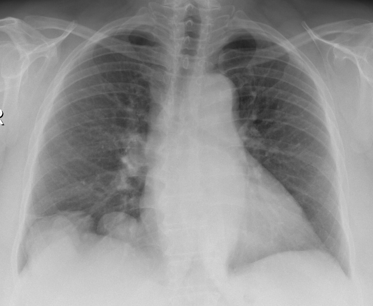
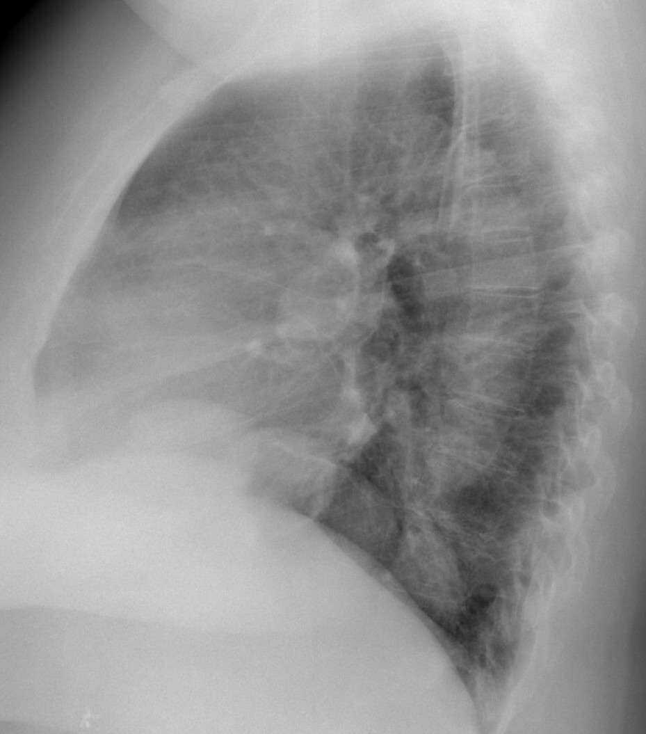
Today I am presenting a case lent to me by my good friend Dr. José Luis López Moreno. The images are preoperative radiographs of a 61-year-old woman with a pelvic mass. What do you see?
Check the images below, leave your thoughts in the comments section and come back on Friday for the answer.
Click here for the answer
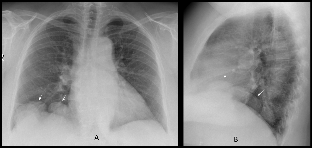
Findings: chest radiographs show several well-defined nodules at the right lung base (A-B, arrows). Considering that the patient has a pelvic mass, a likely diagnosis is metastases from an ovarian carcinoma.
Enhanced CTs of the right upper quadrant show that the nodules are in intimal contact with the liver and have similar density to the liver parenchyma (C-D, arrows). Coronal and sagittal enhanced CT demonstrate liver vessels going into one of the nodules (E-F, red arrows). A follow-up CT six months later (not shown) did not show any change.
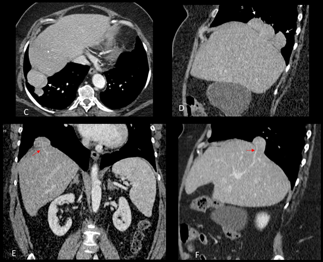
Final diagnosis: multiple liver hernias through the diaphragm.
This case is highly unusual (never seen it before). I did not expect you to make the diagnosis but hoped to be forgiven because of the beauty of the images.
Teaching point: remember that any apparent lung abnormality adjacent to the diaphragm may arise from below, as this case proves.


Pleural based opacities in the right lower lung zone. ? Metastasis from pelvic mass.
Did some research, so, if unusaul, we can exclude pulmonary or pleural mets. Avm doesn’t have any specific correlation with pelvic mass. Endometriosis maybe? Found on radiopaedia if there is lung involment it is almost always on the right side.
You did your research. But remember that to ask the correct question you have to know half of the answer
Malignant Secondaries
Hi,
I’ll check tuberculosis first ( obvius i’m from Algeria)
Then CA 125
They look very unusual for TB. And we also have tuberculosis in Spain
Round opacities could be benign tumor originating from the diaphragm,
pulmonary cysts or hamartomas.
Also cardio-thoracic ratio > 0,5.
greetings,
several large round opacities in the right lower lobe, and probable one in the right hilum, the opacities seem to be pulmonary rather than pleural-based.
mail DDX: metastatic disease, hydatid cysts,..
Right lower lung masses metastasis
Depending on what the pelvic mass is, and previous films, benign metastatizing leiomyoma might be a viable option.
I see multiple opacities- nodules in LRL- some metastasis? And large right hilus?
Sarcodoises
You are all doing well, but so far the right answer has not been mentioned. Go back to the basics
well shaped, high density in the bases and around the hilum. An obese patient.
probably pleural lipoma?
some nodules in soft tisue in right?
I think they silhouette the diaphragm. Could they all be just unusually looking humps? Or maybe something else arising from the diaphragm?
Could they be arising from below the diaphragm?
Liver mets?
Not really, but close. See the answer tomorrow. Well done!
Then either peritoneal implants or pseudomyxoma peritonei from ovarian (mucinous?) carcinoma.
There are several opacities on top of the right diaphragm, with the diagnosis of pelvic mass I’d suggest Mets. Also augmentation of descending aortha caliber, aneurysm.
Intravenous spread of uterine leiomyomatosis to lung