
Dear Friends,
Muppet is getting soft in his old age and presents you with an easy case: pre-op chest in a 40-year-old male with a brain tumour.
Diagnosis:
1. Lung metastases
2. Lung carcinoma
3. Pleural tumour
4. None of the above
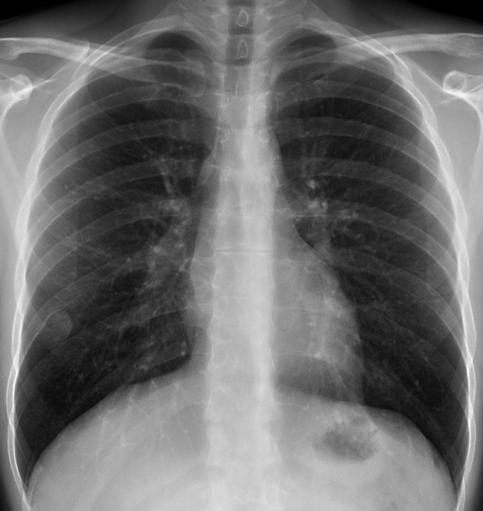
40 year-old man, PA chest
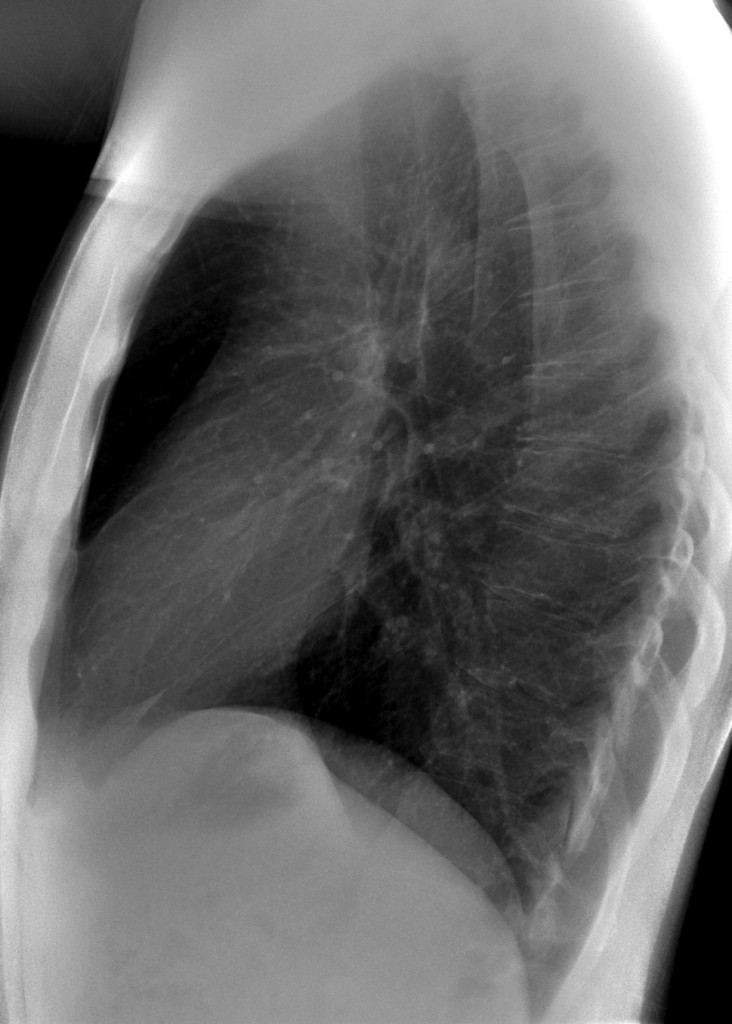
40 year-old man, lateral chest
The PA chest film shows a rounded lesion with the ‘incomplete border sign’ (medial aspect outlined by air, lateral border not visible because in contact with chest wall). In addition, there is erosion of the lower border of the rib (arrow). This combination of signs is pathognomonic of a mass in the underside of the rib. CT confirms the presence of a soft-tissue mass and the erosion of the rib (arrows).
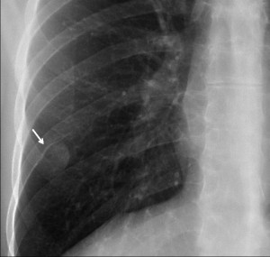
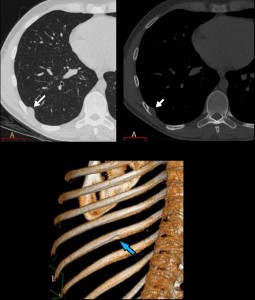
Final diagnosis: neurofibroma. The patient had neurofibromatosis and a meningioma. Muppet is very happy with your diagnostic skills and your semiological interpretation.
Teaching point: when the underside of the rib is eroded, the lesion arises from a structure which travels under the rib: a nerve (as in this case), vein, or artery
(remember the erosion in aortic coarctation).


4. None of the above.
Neurogenic tumour is a good posibility for me. There is an opacity that is associated to a notch at the seventh right rib. There is not destruction of the rib. There are other findings of lesions placed out of the lung in this case: the edge of the opacity is clear at its inner portion. This portion is surounded by lung. But you couldnt say where the edge is in the rest of the lesion (the portion that contacts with the chest wall).
I can´t see the lesion on the lateral view. Pleural tumor is a lesion that grows out of the lung. Neither pleural lesions nor rib metastases usually remodel the bone.
Sorry, I would mean eighth rib.
mmm..i don’t know :/ it can be 3 or 4, there is a small rib deformation in the right side
on the lateral view I think it’s in the Th IX pedicle level
3. Pleural tumor because of incomplete border sign and indenting of the lower border of the postero-lateral arch of VIII-th right rib. Probably benign since there is uniform sclerosis and no cortical interruption at contact.
I totally agree with Lola: the semiology indicates that it is an extrapleural slow-growing lesion. In this location I also would think of a neurogenic tumor as the first possibility.
Totally agree with Lola: the semiology indicates that it is an extrapleural slow-growing lesion. In this location I also would think of a neurogenic tumor as the first possibility.
None of the above
Because it seens like the lesion is producing scalloping of the inferior border of the 8th posterior rib, thus the lesion should be outside of the pleural and pulmonary cavity, and should be located in the thoracic wall/rib.
It could be a pleural lession since I don´t see it in the laterla view, but I´m not sure. Mr muppet, that is not such an easy case! 🙂
It could be a pleural lession since I don´t see it in the lateral view, but I´m not sure. Mr muppet, that is not such an easy case! 🙂
I believe the case is easy if you see the erosion on the underside of the rib. In such case, the two main diagnosis are neurogenic tumor or aneurysm of intercostal artery (much more common the first one)
I think that the origin is pleural for tre reasons and maybe could be benign (lipoma or fibroma).
1) It’s possibile look only the medial border
2) Can’t look in L-L view
3) Make encasement in costal arch without osteolysis
N.4 : t.neurogeno 7 costa.