Dear Friends,
Muppet promises never again to show a bone case. He has been severely reprimanded!
Back to good ol’ chest imaging:
29-year-old male heavy smoker. Admitted with mild dyspnea.
Diagnosis:
1. Mediastinal tumour
2. Dilated cardiomyopathy
3. Pericardial effusion
4. None of the above

29-year-old male, heavy smoker
Click here for the answer to case #21
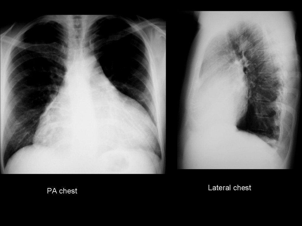
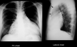
PA chest film shows apparent global enlargement of the cardiac silhouette, with no engorgement of the lung vessels. At first sight, the appearance indicates pericardial effusion. However, the clue to the correct diagnosis lies in the lateral film, which shows occupation of the anterior clear space (arrow), which should be blacker in a 29-year-old person. This finding suggests an anterior mediastinal mass.

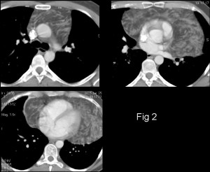
CT confirms the presence of a fatty mass originating in the anterior/superior mediastinum that extends caudally and wraps itself around the heart (fig 2). Thymic masses occasionally grow caudally, appose themselves around the heart and simulate cardiomegaly in the chest radiograph.

Final diagnosis: thymolipoma, surgically proved.
Teaching point: always look at the anterior clear space in the lateral chest radiograph. You may find pathology not visible in the PA radiograph.


1. Mediastinal tumour
Mediastinal tumour and pericardial effusion.
Well, back to chest… bone is no fun- it’s all about experience there; plus no good books under 5-volume-sets.
So i see the X-ray is carefully blackened so as not no see signs of vascular redistribution; or is it so as to show the peculiarly rounded inferior right cardiac border ? Anyway: the heart enlargement seems bottle shaped (but on profile the epicardial fat pad is not displaced), overlaps pulmonary hilli without {visible} pulmonary vessel enlargement. I see also a septal thickening in the left pulmonary base (Kerley B?) but no pleural effusion. I see no signs of cardiac failure. Apex of the heart is ascended and inferior left cardiac arch is rounded. So i won’t go with pericarditis, but with a right heart enlargement (that goes with smoking) + maybe a pericardial cyst ? I’d have a echo than CT to diagnose.
Muppet concerned abot the few answers. Is he losing the favour of his followers? Case too easy/difficult?
Hint: look at the lateral view
Tumor, mass or cyst. Pericardial cyst?
Enlargement of the heart shadow that hides the hila related to pericardial effusion.
Occupation of the retrosternal space on tha lateral view suggesting a possible anterior mediastinal mass.
The pericardial effusion doesn’t allow to asses the hila on tha AP, but on the lateral view is possible that the left pulmory artery shadow was enlarged and nodular (especially in its craneal part where seems to be two nodular spiculated opacities). Then also seems to exist occupation of subcarinal space giving the “donut”sign. That suggest extensive adenopathies.
I propose lymphoma (anterior mediastinal mass and extensive adenopathies) with pericardial effusion.
Better late than never! But Muppet disagrees with some parts of your comment. Other that the shape of the cardiac shadow (unreliable finding) you don’t have any criteria to diagnose pericardial affusion.
Aside from this, good discussion
Globular shape enlarged cardiac shadow,pericardial effusion.
Loss of retrosternal Lucency likely mass in the anterior mediastinum.
Opacity with irregular margins in the upper zone.
Hyper translucent lung fields.
A diagnosis Anterior mediastinal mass cannot be challanged. However possibility of spring water cyst should also be considered. Lateral view clearly shows anterior mediastinal opacity. Further evaluation with CT thorax needed.
La silueta cardiomediastínica está muy aumentada en la proyección PA y en la lateral se observa una ocupación del espacio retroesternal, lo que me sugiere una ocupación del mediastino anterior. Diagnóstico diferencial: 4 Ts….
There are only 3 Ts: thymoma, teratoma and “terrible lymphoma” The fourth T (thyroid) belongs in the superior middle mediastinum. All according to the Muppet, who is nearly infallible!
Good discussion. Muppet awed
I agree thyroid belongs to the middle mediastinum most commonly, but i wouldn´t exclude it from the anterior mediastinum.
PS: finally i got the right answer!! mediastinal tumor! 🙂
Yes, you did. Congratulations!
1) Globular heart shadow occupying more than half of the thorax, goes with pericardial effusion.
2) loss of normal anterior mediastinal lucency, yes it could be lymph nodes but then the frontal view doesnt support it much ?
3) lateral view shows abnormal density projected over the heart shadow , again raising the possibility of a mass.
4) lungs clear, TB less likely
so i’ll go with pericardial effusion. second possibility is dialated cardiomyppathy.
fingers crossed and WAITING for muppet to break the shackles !
Sorry, Muppet walked close to kryptonite, shackles will stay for a while longer.
You are in for a dissapointment, though.It’s not what you think. Answer coming soon.
Mediastinal lipomatosia
rodis iqneba swori pasuxi?
mediastinal lipoma/liposarcoma
I posted the answer and nobody considered it!
Sorry. Muppet recognizes your contribution. But it will be unfair to the other participants to give away the answer too early. From now on, when anybody makes the right diagnosis Muppet will tell him/her on the private mail.
looking only at the pa film I would have started the differential diagnosis for an enlarged heart. interestingly on the lateral film, there is opacification of the retrosternal space indicating an anterior mediastinal mass. 3 Ts for diagnosis, I agree.
any signs on the pa that indicate that this is not enlarged heart?
No signs in PA. Lateral view helps to diagnose mediastinal masses and pericardial fluid
I think it is a mediastinal Tumor (for example: Thymoma or Thymic Caner).
Iam not sure, but i think you showed us a similar case in the lecture of Mediastinum in ECR2012 🙂
Yes, I did. Good memory!
nON è UNA MIOCARDIOPATIA Nè UN VERSAMENTO PERICARDICO, PERCHè LO SPAZIO RETRO-CARDIACO è LIBERO( VEDI L.L.)E NON CISONO SEGNI DI SOFFERENZA DEL PICCOLO CIRCOLO, TRANNE UN PICCOLO VERSAMENTO PLUERICO NEL SENO COSTO-FRENICO SX. nON è UN TUMORE MEDIASTINICO PERCHè LA LA MASSA ABREBBE CONTORNI IRREGOLARI E CI SAREBBE UNA CLINICA DI INGOMBRO MEDIASTINICO. SAREBBE ALLORA LA 4 IPOTESI DI UNA PSEUDO-MASSA A PARTENZA DAL MEDIASTINO ANTERO-SUPERIORE CHE IN L.L. è OPAco: penso allora ad una cisti timica( la TC ne confermerebbe il contenuto liquido)
Thymoma for me would be the first diagnosis subjectively there is a slight wavy but how does smoking fit in? May be incidental?
Pericardial effusion.
Podria ser Cardiomiopatía dilatada, garcias al consumo exesivo de sustancias toxicas, quimicas y peticidas que contiene el tabaco
Pericardial cyst???? 4ts (CT is needed…)
endocardatis fommowing pneumonia?
3 Ts or mediastinal fat.
Illustre collega come vedi dal mio commento,ho indovinato la patologia come di pertinenza timica: non si poteva dewfinirne il contenuto se non con la TC (cisti timica v-s timolipoma):non pensi allora che sia stato bravo? Perchè non ha commentato la mia risposta? Non conosco ben l’inglese ecco perchè mi sono espresso in italiano!
Sorry. No problem with writing in Italian. Muppet gets by in several lenguages.
Muppet congratulates you for being right
Grazie per le tue belle parole!!!!!