Muppet is feeling good today (love letter from Miss Piggy) and wants to show you an easy case: a 69-year-old male with chest pain and productive cough. TB in his youth.
1. Active TB
2. Empyema necessitatis
3. Pleural abscess
4. None of the above
Dear friends,
I don’t believe that the case is difficult. In fact, if you analyse all the findings, it is reasonably easy.
The chest radiograph (TB in youth) shows signs of chronic pleural disease: small hemithorax and calcified pleural thickening.
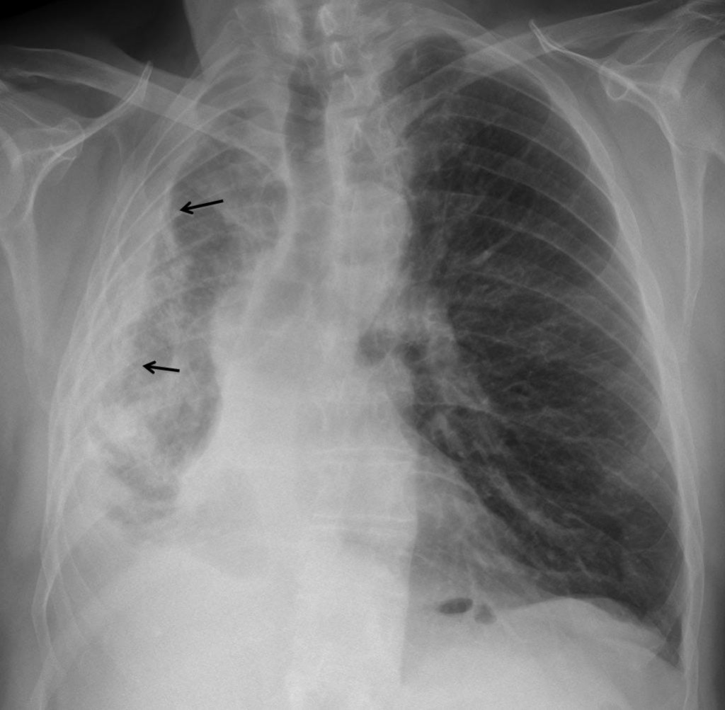
Fig. 1
CT confirms the findings and shows a solid component (arrows) with invasion of the chest wall (arrows).
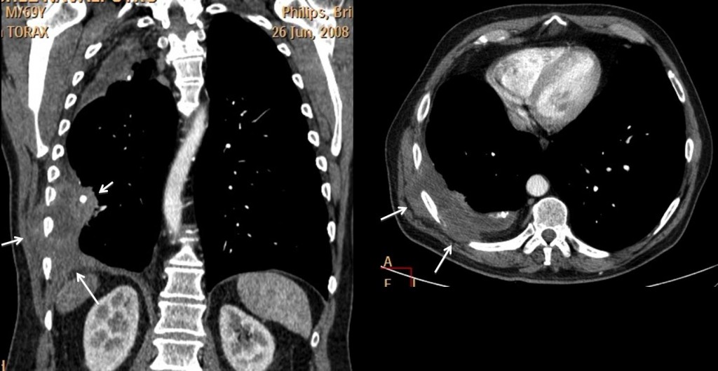
Fig. 2
This combination is highly suggestive of malignancy developing in chronic pleural disease (lymphoma, carcinoma or mesothelioma. They all look the same). Empyema necessitatis can be discarded because it is mainly fluid and has no solid component.
Final diagnosis: lymphoma developing in chronic pleural disease. Patient is alive and well four years after initial diagnosis. Ref. AJR 194:76-84; 2010
Teaching point: complications of chronic pleural disease are mainly three: bronchopleural fistula, empyema necessitatis and malignant tumour. CT can easily distinguish between the last two, because of the solid component of the tumour.

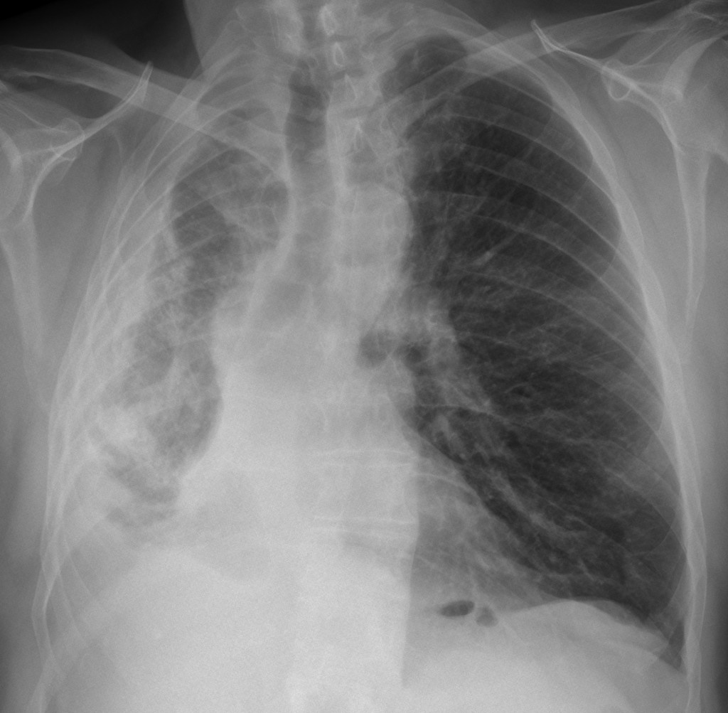
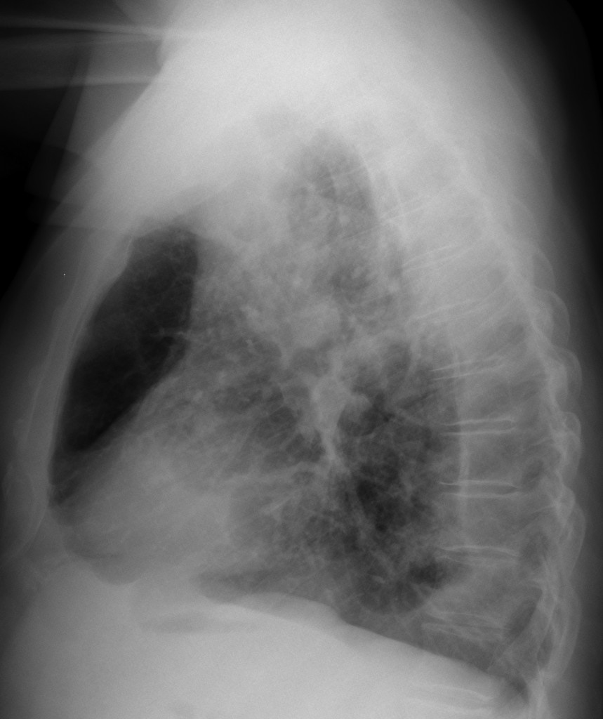
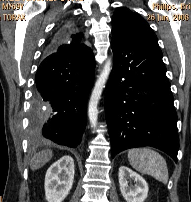
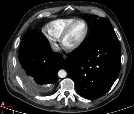


Heterogenous pleural thickening with lower attenuated areas, calcifications and bulging from intercostal spaces. Narrowed intercostal spaces in the right hemithorax compatible with decreased right lung volume secondary to pleural disease. No bone destruction adjacent to pleural lesions.
My first choice of answer is empyema necessitatis.
4. Mesothelioma
Mesothelioma
there is external extension superfiscially is seen so i in favor of no 2 . Empyema necessitatis
Il caso sembra abbastanza facile(oppure l’illustre collega ha fatto pre-tattica?).Ipoespanso e velato l’emitorace dx ,con attrazione omolaterale dell’ombra cardio-mediastinica, all’Rx-stadard.La TC, con mdc, conferma tali dati ed “aggiunge”, calcificazioni pleuriche, sospinte internamente da versamento che appare in taluni tratti, piu’ipodenso, come da componente liquida:non c’è CE pleurico ed esternamente vi è “tumefazione” delle parti molli con le stesse caratteristice. CD Empiema necessitatis( numero 2).
Heterogenous pleural thickening with calcifications and collections is seen in the left pleural cavity. There is extension into the soft tissues of the chest wall. I go for empyema necessitatis.
2.empyema necessitatis
I cannot tell between mesothelioma and empyema necessitatis
Yes, you can
Decreased right hemithorax volume and pleural calcifications. No mesothoracic pleural lobular thickening. The ribs are not destroyed, instead there is bulging of the hypoattenuating lesion through them. I think this is empyema necessitatis.
Empyema necessitatis.
Plain CXR: Mediastinal shift towards the right. Right hemithorax demonstrates decreased expansion. Pleural thickening and blunting of the right costophrenic recess noted.
Lateral CXR does not demonstrate lobar collapse/consolidation.
CT coronal reformats and axial imaging demonstrate calcified pleural thickening and a collection with areas of hypoattenuation extending across the chest wall in keeping with EMPYEMA NECESSITANS.
Empyema necessitatis
The PA and lateral view CXR shows extensive pleural reaction along the lateral and posterior chest walls in right hemithorax with flattening of the right dome.Few specks of calcification are seen along the medial aspect of the thickened pleura in right upper zone. There is evidence of volume loss on right side in the form of overcrowding of the ribs, tracheal and mediastinal shift to the right and scoliosis. Interstitial thickening with fibrosis is seen in the right lung with compensatory hyperinflation of the left lung. No rib destruction is seen on right side. Small air lucencies are seen in the retrocardiac location on left side(may represent incidental hiatus hernia). The x-ray features are suggestive of chronic pleural pathology with fibrothorax on right side-due to tb(previous history of tb present). The other differentials will include chronic empyema(due to other infective causes),mesothelioma or asbestos related disease and chronic resolved hemothorax. Coronal and axial CT sections confirm extensive pleural thickening with some low attenuation areas within it extending along the intercosal space into the soft tissues of the right lateral chest wall without obvious rib destruction. Extraplueral fat is also thickened confirming chronic process. The diagnosis based on history and imaging findings is empyema necessitans. the differentials like chronic hemothorax can be excluded based on no history of trauma,no rib fractures. Features in favor of mesothelioma are pleural calcification(may represent calcified pleural plaques -not typical site for plaques-they are typically seen along the posterior pleura along lower chest wall and along diaphragmatic pleura) and pleural thickening more than 1 cm but mesotheloima can be excluded in this patient as no history of asbestosis, no circumferential(or rind like) pleural involvement and no involvement of the mediastinal pleura.
Empyema necessitatis
Empyema necessitans, for the reasons outlined above by colleagues.
Why do you think your colleagues are right?
This is not mesothelioma – there is no nodular cirumferential pleural thickening encasing the lung, no pleural effusion. And mesothelioma does not usually extend in the soft tissues…
Besides that, unilateral pleural calcifications make me think of previous TB empyema or previous haemothorax…not asbestos exposure, where such calcifications would be bilateral.
Do you agree Dr Pepe?
I have seen mesotheliomas invading the ribs occasionally. I agree with you about the unilateral calcifications going against asbestos. But there why, if it is a tumor, has to be a mesothelioma?
Well, this is not intrapulmonary…neither does it look like an extrapleural process…it looks more like an intrapleural process invading the soft tissues.
I did not say that mesotheliomas don’t invade/destroy the ribs – anything is possibly. What I said is that it is rare for mesotheliomas to extend in the soft tissues.
The only other tumour I can think of is pleural fibroma…the imaging features highlighted here are not those of fibroma.
Them you can have lymphoma, mets, and occasionally extension of a thymic tumour. Then again, it does not look like it.
The unilateral pleural calcifications (? previous tb empyema/previous haemothorax), the apparent low density content of this abnormality, and the invasion of the soft tissues make me favour empyema necessitans.
Mesothelioma
Reading your comments to the chosen diagnoses above makes me think there is more to this case then one would like to think…
There seems to be a nodular mass above the right hemidiaphragm (coronal ct), plus the above mentioned pleural fluid collection that invades the thoracic
wall.
I have only seen mesothelioma 2-3 times and empyema
necessitatis once so far; mesothelioma consisted of circumferential nodular pleural thickening and multiple lung nodules.
There was stronger enhancement around the fluid collections in Empyema necessitatis & the Patient was very sick.
I would like to know more of the anamnesis and the blood work results.
However malignancy must be ruled out (carcinoma on top of chronic pleural disease post-TB)? Thanks for another challenging case, it can be frustrating though – Radiology is so very difficult.
You are very close. Don’t despair. Review last two years of AJR.
Il Real-Madrid ( Muppet) ha fatto pretattica(contro il Bari): il caso infatti NON è facile! Poi, si è impetosito contro una squadra di serie B e le ha dato l’aiutino ( AJR):ianuary 2010;194:76-84. Diagnosi finale:Pyotorax-associated Lynphoma!
As you can see, the case was easy. You even have a reference! Congratulations.