Dear Friends,
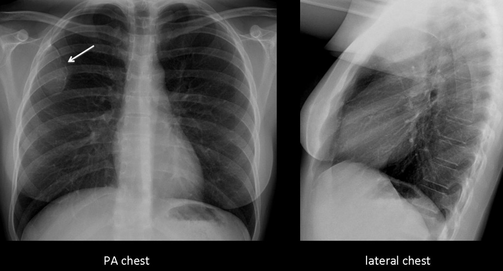
Since you are getting used to difficult cases, Muppet is showing you an easy one. May the Force be with you. It is a pre-op chest radiograph of a 37-year-old woman with breast carcinoma.
Diagnosis:
1. Metastases
2. Granuloma TB
3. Hydatid cyst
4. None of the above

37-year-old woman, PA chest

37-year-old woman, lateral chest
Click here for the answer to case #36
Findings: the PA chest film shows a solitary expansive lytic lesion in the anterior aspect of the third right rib (arrow), without an accompanying soft tissue mass.

Fig. 1
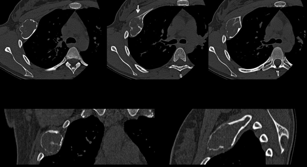
CT confirms the findings and the lack of soft tissue component. There is cortical thinning and a short transition zone with normal bone (arrow). The appearance of the lesion is very suggestive of a benign process and, as many of you pointed out, fibrous dysplasia is the first possibility.

Fig. 2
Final diagnosis: fibrous dysplasia.
Teaching point: Solitary expansive lesion in a rib in a young person is highly indicative of fibrous dysplasia, which is the most common benign lesion of ribs.


here it goes… well-defined expansile lesion of the 3rd right rib anteriorly – ground glass matrix – this is most in keeping with fibrous dysplasia (renal cell carcinoma may characteristically give expansile lytic bone mets but breast cancer gives mixed lytic/sclerotic.)
Answer D: NONE OF THE ABOVE
Agree with fibrous dysplasia – other considerations are enchondroma, though less likely due to the appearance.
In older patients, we should also consider plasmacytoma and metastases.
non of the above
begnin boney lesion
Chest x-ray showing expansile lytic lesion involving the right 3rd rib anteriorly. no obvious soft tissue component seen in relation to it. other bones are normal. No lung lesions seen. cardiovascular shadow is normal. Soft tissue shadows are normal. The thoracic vertebrae are normal. no rib notching seen. The differential diagnosis of this apperance in an adult include-fibrous dysplasia,mets from RCC/thyroid, plasmacytoma, brown´s tumor, chondrosarcoma, paget´s disease.Diagnosis is fibrous dysplasia(young age and ground glass matrix, typical location). Mets from RCC and thyroid can be excluded on the basis of no previous history,no lung lesions, only single rib involvement(though it can occur),expansile lytic mets are not seen in breast CA, brown´s tumor in rib can be excluded on the basis of no other changes of hyperparathyroidism(like rib notching, rugger jersey spine, no history of chronic renal disease,normal visualised clavicles). Age is against paget’s and chondrosarcoma(also no arc or ring like calcifications seen within the lesion). Enchondroma and plasmacytoma are strong D/D.
I go with fibrous displasia in the anterior arch of 3rd rib.
None of the above
Beware the muppet! He can be evil.
Remember that Dr. Cáceres works in a mediterranean tertiary center, so I’ll go with Hydatid cyst.
non of the above
begnin boney lesion
Lesione espansiva dell’arco anteriore della 3 costa con caratteri semeiologici di Benignità( si esclude la metastasi).L’interessamento di una TBC, si può ragionevolmente escludere dalla negatività polmonare ed ilo-mediastinica.La cisti da echinoicocco dovrebbe avere un cercine osseo piu’ marcato.Rimane la osteolisi intracorticale, che”soffia” l’osso assotigliandone la corticale, come nella Displasia fibrosa , monostotica(escluderei l’encondroma, per la mancanza di evidenti tralci ossei intratumorali).
benign tumor of the 3th-right rib
Expansive lesion is identified and low density defined ranges, which seems to depend on the portion of the third rib axillary right. It is suggestive of monostotic fibrous displasia.
congratulations for the writing. lista de emails lista de emails lista de emails lista de emails lista de emails
you must read cheap ugg with low price