Muppet is impressed with your knowledge. He tries very hard to post teaching cases and has decided to skip the multiple-choice questions in the following chest case, which is that of an asymptomatic 56-year-old male.
1. Where is the lesion?
2. What is your diagnosis?
The first five radiologists to suggest the correct diagnosis will be given a DVD at the next European Congress of Radiology.
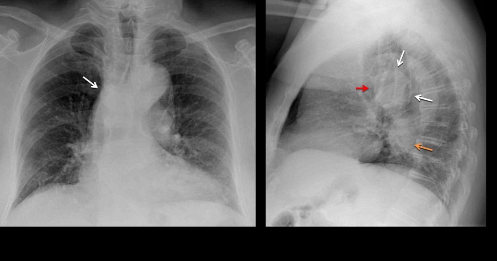
Chest radiographs show a rounded opacity in the middle mediastinum (white arrows), imprinting in the lower trachea (
red arrow). There is a lower rounded opacity (
orange arrow), which represents the tortuous descending aorta.

Fig. 1
Answering the first question, the abnormality is located in the middle third of the middle mediastinum. Most common masses in this location are lymph nodes and congenital duplication cysts. However, the location in the PA view and the imprint in the lower trachea should suggest an aberrant left pulmonary artery (pulmonary sling), as Raffaela mentioned.

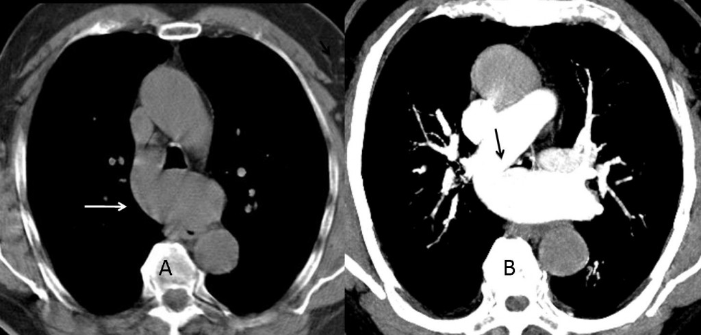
Fig. 2
CT confirms the diagnosis by showing the anomalous artery (A, arrow) crossing between the oesophagus and trachea and the origin of the left pulmonary artery from the right (B, arrow).
Diagnosis: aberrant left pulmonary artery (pulmonary sling).
Congratulations to Raffaela for her excellent diagnosis. You can choose from the Godfather trilogy, The Lord of the Rings, or The Bourne trilogy.
Teaching point: always look at the trachea in the lateral film. It is an excellent marker for discovering middle mediastinal masses.


La patologia è nel mediastino medio.Il calibro della trachea è aumentato.Diagnosi: S. di mounier-kohun( tracheo-bronco megalia)
Arch of aorta & cardiomegaly
There is evidence of a linear radioopacity silhouetting the right heart border. The right hilum is inferiorly displaced. There is bilateral hilar and supra hilar adnopathy more evident on lateral film as donut sign. Hence, an enlarged lymph node is compressing the right lower lobe bronchus causing collapse.
The esophagus appears to be dilated. No definite air fluid level in it or mottled lucencies. No rib resection. No sign of aspiration. Difficult to appreciate gastric bubble on PA view, I feel it is present on lat view.
Mild cardiomegaly. Age related unfolding of aorta and degenerative changes in spine.
To conclude, malignant lesion of esophagus with metastatic hilar lymphadenopathy causing partial collapse of the right lower lobe.
I would strongly suggest CT Chest with IV contrast for further evaluation.
CT will be shown in due time. It’s more fun to suggest the diagnosis in the plain film
1.Trachea and mainstem bronchi are dilated and I think there are some upper and lower right lobe bronchiectasis.
2.Mounier Kuhn syndrome
ascending aorta aneurysm?
The lesion is in the posterior mediastinum. On the lateral view i see increased size of a neural foramen. I think it is a neurogenic tumor
Tracheal diameter is dilated, more evidently in the lateral proyection suggesting Mounier-Kuhn Syndrome
Posterior basal segment right lung.
Lung lesion.
On the posteroanterior chest radiograph I can’t appreciate the azygoesophageal line thus, in my opinion, the abnormality can be located in both the middle and posterior compartments.
Nevertheless, some more informations are given us by the lateral chest radiograph in which we can see abnormal dilatation of the esophagus…so could it be achalasia?
1.posterior mediastinum
2.Esophageal diverticulum
Double shadow at the aortic knuckle
Aortic anurysem
PA and lateral chest x-ray shows ill defined focal lesion in the retrocardiac location on lateral view overlapping the aortic shadow. No calcification or cavitation is seen within it. prominent vascular markings are also seen adjacent to it. The lesion appears to be in right lung . right hilum is also lower in position as compared to left. no evidence of any loss of lung volume is seen. Left pulmonary conus is prominent-vascular/?lymphnodes.aorta and left heart border are normal. Right heart border is not well seen.no pleural thickening or effusion seen. Right lung lesion-the differentials include primary lung neoplasm, sequestration,round atelectasis. there is also suspicion of a pseudonodule in the left midzone in PA view overlapping the anterior aspect of left 4th rib.
– On the basis of the PA e LL projections, the abnormality is located in the middle mediastinum and, in my opinion, this is a case of anomalous left pulmonary artery (pulmonary artery sling), a condition that can be asymptomatic in adult.
– To support my thesis: on frontal view there is an opacity at the right tracheobronchial angle which is seen behind the trachea in the lateral view.
Good explanation. I think you have the correct the diagnosis !
Su suggerimento di quanto evidenziato da alcuni collego, rivedo la mia posizione( sempre convinto che l’immagine tracheale sia allargata). Opacità a banda , in sede paracardiaca basale dx, con scarsa evidenza dell’ilo (in AP).In laterale opacità triangoliforme , nel mediastino medio, con apice all’ilo e base su diaframma.Atelettasia del lobo medio,con linfoadenpatie(in LL), come da S. del lobo medio.
I know that I said this already – but I think that image quality affords to be better…in real life we get better images. We could even get much better image quality if the JPEG file is saved in the least compressed format, or if there is an option to download TIFF files for each case and view them on our PC.
Having said that, I would like to thank all the people behind this European joint effort!
Will convey your suggestions to the technical staff. Hope you will agree with me that the imprint in the posterior inferior wall of the trachea is very obvious. Sometimes, in real life, quality of films is not excellent and you have to do what you can.
Thank you for the compliment.
Rafaella is great!
Her diagnosis also explain the volume loss of the right lung -i.e. chronic bronchial compression.
Pulmonary slim.
Yes, I agree with you that Raffaela gave a good opinion.
In the PA , I see some nodular structures projected behind the heart in the left hemithorax. In the Lateral view I still see them, some of them projected on the heart and the other projected in the Inferior hemithorax. I do not know If this is real, and could be related with some kind of anomaly in the left pulmonary veins dreinage.
But I also agree that the explanation of the Aberrant Left pulmonary artery may has sense.
Sempre convinto della tracheo-megalia, essa si spiega con il suggerimento della Raffaella, come da impronta vascolare sulla parete posteriore inferiore della trachea( da anomalia di decorso dell’arteria polonare sx).
This case is breaking my head.
The main findings that I find are:
-Middle mediastinum mass (I have been thinking of multiple vascular variants but no one has convinced me at all).
-Lineal sclerosis of the vertebral endplates.
-Osteolisis in posterior arch of the 8th right rib.
Only one option to explain all of them came to my head.
I think it could be an ectopic mediastinal functioning parathyroid adenoma causing some of the bone changes of hyperparatiroidism (rugger jersey spine and the osteolisi on the right rib wich coul even be a brown tumor).
It’s a bit crazy but I have no more ideas.
pulmonary artery sling ! apparent on frontal projection above the right hilum and on lat projection, between the inferior aspect of the posterior wall of trachea and anterior wall of the esophagus.
Aortic aneurism.
Dr.Caceres:
Wonderful case. I wonder about the apparent dilation of the left PA. Is this a usual manifestation of the condition?
Many thanks.
Herb Kaufman
To tell you the truth, I don’t know. I have seen only three cases in adults and don’t remember the other two having a large left PA. Let’s hope that somebody else has a larger series and answer your question.
Thank you
check chanel outlet for gift