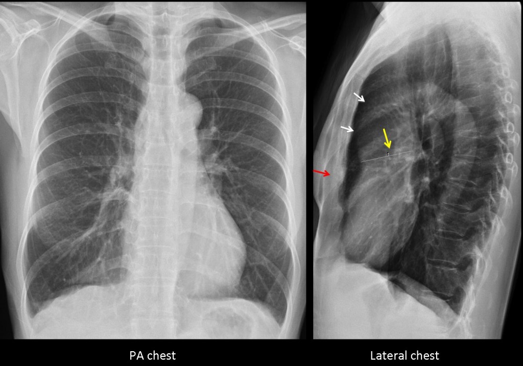
Since we had very few answers in the previous case (too easy or too difficult?), Muppet is reverting to plain films and straightforward questions. The following are pre-employment radiographs of a 43-year-old female. She was told that she had enlargement of the ascending aorta. What would be your diagnosis?
1. Marfan’s
2. Aortic coarctation
3. Aortic valvular disease.
4. None of the above
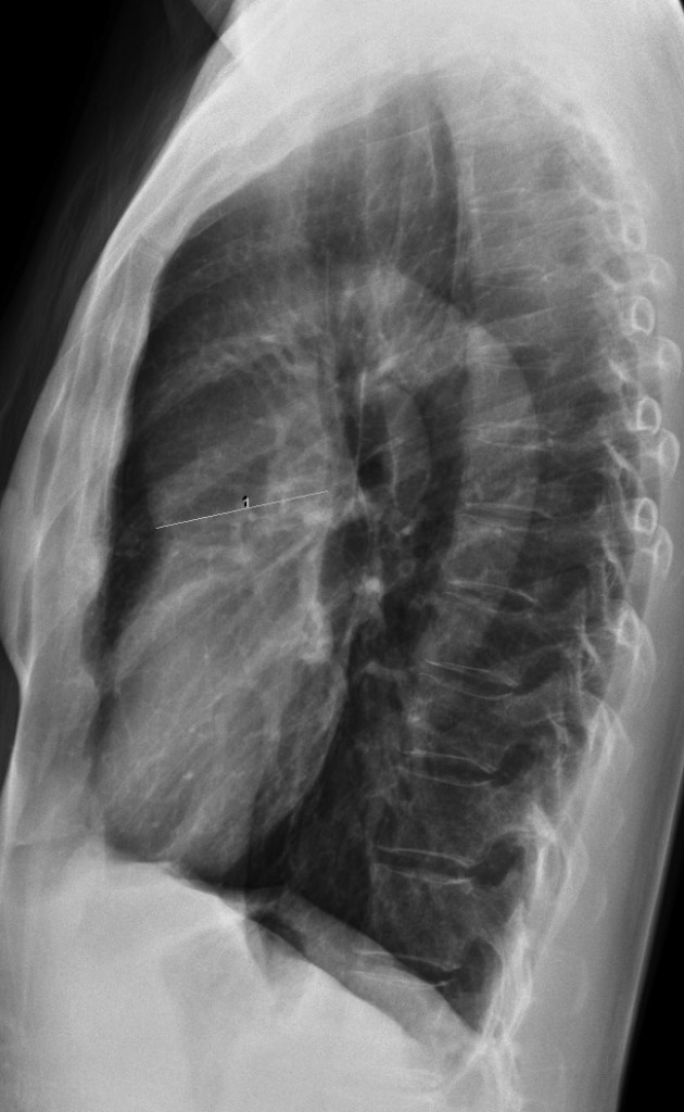
Findings: PA chest film is unremarkable. The ascending aorta looks normal. Lateral film shows a prominent ascending aorta (white arrows). A line has been traced which measures an apparent dilatation of the ascending aorta (yellow arrow). The obvious error is that the line is drawn from the anterior aspect of the aorta, outlined by lung air, to an imaginary point in the mediastinum, which may or may not be the posterior aortic wall. It is important to remember that the inner wall of the aorta contacts with the mediastinal tissues and, according to the silhouette sign, cannot be visible. Therefore, whoever traced the line is wrong.
MRI confirmed that the aorta was normal and that no other abnormalities were present. The apparent prominence of the ascending aorta in the lateral film is due to a combination of a moderate pectus (red arrow) and a narrow AP diameter of the chest.

Fig. 1
The patient is friend of mine and the examination was made in a university hospital abroad. MRI is not shown because I could not obtain it.
Final diagnosis: normal chest
Teaching point: do not forget that in the chest radiograph we only see the outer contour of the aorta. It is impossible to see the inner wall because it blends with mediastinal structures. Remembering this basic principle will avoid errors like the one that happened in this patient.
And never, never, draw lines in the radiographs!


There is knotching of the ribs,that suggesting an aortic coartation.however 40year old age can be considered a late diagnosis,and patient should have symptoms.
Patient is asymptomatic
i think non of above suggestions…..another diagnose
😀 😀 😀
ascending aortic aneurysm
pseudocoarctation?
It’s Marfan’s syndrome, causing ascending aortic aneurism.
Ascending aortic aneurism. The woman seems to be tall and thin. Pectus excavatum. No scoliosis.
I cannot see the rib-knotching.
Aortic valvular disease should be sympthomatic.
On balance, it might be Marfan’s… or a thin woman with aortic aneurism.
There is dilatation of the aortic root and the ascending aorta is aneurysmal. There is some narrowing at the junction of the aortic arch and the descending aorta suggestive of post-ductal coarcation of the aorta, possibly with a congenitally bicuspid aortic valve.
Differential diagnosis includes pseudocoarctation, aortic stenosis with post-stenotic dilatation, Ehlers-Danlos syndrome, Marfan’s syndrome and homocystinuria.
coarctation aortique
Pseudocoartation the final answer should be thebdiagnosis
The most likely diagnosis is the aortic valvular disease, because:
– the enlargement of the ascending aorta in a supravalvular location seen on the lateral chest X-Ray
– the convex middle mediastinal arch on the right, from the ascending aorta that pushes a little the SVC at this level, on the PA chest x-Ray;
-the left inferior mediasinal arch is convex suggesting a hipertrophy of the left ventricle
– I cannot see any skeletal abnormalities
– the patient breathed in strongly and we can see the diaphragms are flattened and ribbs are horizontally ;
– I can see the aortic knob so there is no aortic coarctation
– the pulmonary vessels are clearly seen on both sides with a normal pattern.
– I cannot see any calcifications of the aortic valve and the patient is middle aged so maybe the aortic valve stenosis is rheumatic
Findings
dilatation of the aortic root
the ascending aorta is aneurysmal
There is narrowing at the junction of the aortic arch and the descending aorta suggestive of coarcation of the aorta
I cannot see rib-knotching.
The most suitable diagnosis could be aortic coarcation
İf there is not Ribknotching,the aortic coartation is not a good choice,
Muppet agrees with you
TheRe is diffuse dilatation of the asendan aorta,and may be the root of the aorta,.the patient is female and Middle aged,suggesting that an Marfan syndrome.
Nel radiogramma AP, si nota dilatazione del 1 arco di sx(arco aortico);non ci sono anomalie costali e-o vertebrali. In LL si conferma la dilatazione dell’arco aortico ma sopratutto dell’aorta ascendente alla radice;vi è un restringimento(?) nel tratto istmico dell’aorta.La patologia piu’ probabile( la paziente è asintomatica) è una vavola aortica bicuspide, che nel suo decorso naturale può complicarsi con dilatazione della base aortica e con eventuale dissezione cronica.Non mi spiego però il restringimento post-istmico(pseudo-coartazione?).
Ascending aorta anuerysm – no ribs notching and asymptomatic – I’d think about Marfan syndrome, and I’d check eyes, skins etc.
So far, nobody has mentioned the correct diagnosis. Muppet very concerned!
The most striking feature on the PA radiograph is the abnormal aorto-pulmonary window…the aorta on the PA view appears normal, but the pulmonary trunk does not show the normal appearance (on the PA view).
Moving on the lateral view. The relatively well defined structure projecting in the anterior mediastinum is too anterior to be the ascending aorta…in my opinion this is more likely to represent a distended pulmonary trunk. This might be idiopathic and asymmptomatic in young(ish) females…I cannot see any CXR signs of pulmonary hypertension. Other considerations would be post-stenotic dilatation in RVOT stenoses, left to right shunts and anomalous pulmonary venous return – however you would expect some symptoms in these conditions at the given age.
So my final diagnosis is idiopathic asymmptomatic dilatation of the pulmonary trunk.
Now all this is coming from a guy who’s interest is hepatobiliary radiology…so please bear with me if it’s all nonsense! 🙂
You analize the chest film very well, although your final diagnosis is erroneous (see note to Genchi Bari below)
Thanks
It could be Ehlers-Danlos syndrome – but patient is asymptomatic and I wouldn’t make diagnosis based on single Xray.
There is a big list on differential diagnosis of widening of aorta.
Maybe some clues?more informations about patient?
Quello che è strano è che in AP, non c’è evidenza della cosiddetta dilatazione dell’aorta ascendente: questa ipotesi deriva dalla visione in LL.E se foose una immagine di sovrapposizione ? Chiaramente la TC risolve il caso: caro professore voi come avete fatto la diagnosi?
Quella sarebbe una altra possibilità – detto questo sei d’accordo con me a proposito della finestra aorto-polmonare?
SI, in LL non si riesce a vedere lo spazio chiaro della finestra aorta-polmonare
Una dilatazione idiopatica del tronco della polmonare, potrebbe essere diagnosticata con una ecocardiografia ed RM.Dilatazione idiopatica tronco polmonare:BJR (2002)75,532-535.
You thinking goes in the right direction. I saw the films after the patient was told she had an abnormal ascending aorta. I thought the chest was normal.
MRI confirmed that no abnormality was present. Will explain on Tuesday.
Patent PDA ?
Noe of the above
straight back syndrome?
Yes, you can call it that. Congratulations. Muppet elated!
Grazie professore: sei Stellare come il tuo Real !!!!!Bellissima lezione!!!!!
Thank you for the compliment. Although I have to comment that Barcelona has more stars than Real Madrid!
Anche se non ho centrato la….porta!!!!
Excelente caso.Gracias por la enseñanza recibida.