Dear Friends,
After the trepidation of case 42, Muppet hopes you get this case easily. Sixty-eight-year-old male with pain in the chest.
Diagnosis:
1. Tuberculosis
2. Bronchoalveolar carcinoma
3. Actinomycosis
4. None of the above
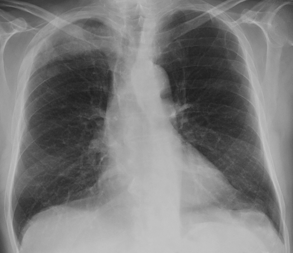
68-year-old male, PA chest
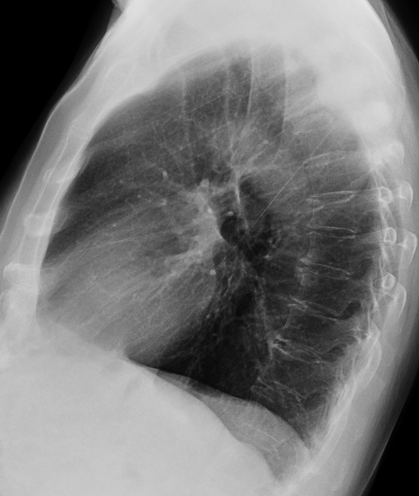
68-year-old male, lateral chest
Post your thoughts and diagnosis below and check back next Tuesday for the answer.
Click here for the answer to Case 43
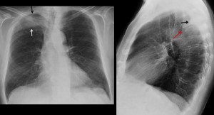
Findings: PA chest radiograph shows an ill-defined opacity in the right apex (arrows). Careful examination shows that the third rib is missing. On the lateral view there is a hint of an extrapulmonary lesion (arrow) and collapse of one of the upper vertebrae (red arrow).
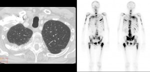
CT confirms the rib lesion, with a significant soft-tissue mass (arrows), which creates the illusion of a pulmonary infiltrate in the chest radiograph. Bone scan shows multiple areas of bone involvement.
Final diagnosis: bone metastases from prostate carcinoma.
From RSNA 2012 in Chicago, Muppet congratulates all of you who made the right diagnosis, especially Dr. Genchi Bari, who made some astute suggestions leading to the correct diagnosis
Teaching point: rib lesions may occasionally simulate pulmonary infiltrates. Always look at the ribs and you may be rewarded.


there is evidence of collapse of D4 vertebra with paraverterbal soft tissue formation.
Also right upper zone shows areas of patchy consolidation.
probably, Tuberculosis..
This x-ray image shows homogenous mass in the upper dexter pulmonary lobe,with retraction of the thorax and mediastinum,but no reaction of the chylus,or lytic lesions of the ribs.We can se also pleural adhaesions in the anterior costo-diaphragmalis sinus.I think the correct answer is none of the above.May be pleural or mediastinal mass.
What about the 4th rib ? Pacoast tumor usually looks like this.
osteolysis of the dorsal part of the 4th right rib with soft tissue component.
A suspiciously denser, nodular area in the dorsal part of the 6th right rib
Also fracture of the 4th left rib of Th4
So i would go for none of the above – either primary or secondary(probably) malignat lesion of the 4th right rib
A me sembra che le lesioni siano tutte ossee: 1 2 3 costa di dx, forse arco anteriore della 6 e lesione dell4 vertebra toracica. Proporrei allora oltre al dosaggio del PSA , totale, e libero, quello del PAP ( fosfatasi acida prostatica) e completerai, se positivi, con una scintigrafia ossea.Mi sembra ovvio a cosa penso.
Easily?
I think the lesions have a skeletal origin.
A dorsal vertebra is collapsed. The right apical opacity could be due to paraspinal abscess or mass in soft tissues and extrapleural space. There is a left paravertebral geographic calcification.
There is also a right upper pulmonary nodule of unknown origin, but I can’t see pulmonary infiltrates.
My diagnosis is 1. Tuberculous spondylitis with paraspinal abscess
N4. I believe there is a mediastinal mass
Looking the lateral X-ray, there is a loss of the normal configuration of the posterior aspect of the 3rd, 4th and 5th right ribs (easily recognised when comparing with the normal ribs caudally). The destroy is more obvious in the 4th rib, when looking the PA film.
Also there is loss of height of the 4th vertebra.
There are no signs of intrapulmonary lesions. Therefore I would exclude answers 1,2,3.
Considering the patient’s age, when seeing osseous destructions, metastasis should be the first diagnostic thought. D/d: multiple myeloma.
(At first I thought of another entity, the Gorham’s disease-because of the continuity of the affected bones, but I excluded it for two reasons: 1st there is no right pleural effusion and second it is to rare to be true!)
there is a lythic lesion of 4th rib and in lateral view there isn’t evidendence of parechimal desease but a convex focal density of pleural/costal compartiment.
I suggest a lesion of chest wall and actinomycosis should be correct. in second istance a costal metastasis.
Il Bari ha vinto le ultime due partite per 3 a O e 2 a O: ammira il mtico REAL ,,,, ( PUSKAS,DI STEFANO, GENTO, anni 50-60), ma non arriverà mai così in alto come il celebre Collega!!!!