
Dear friends,
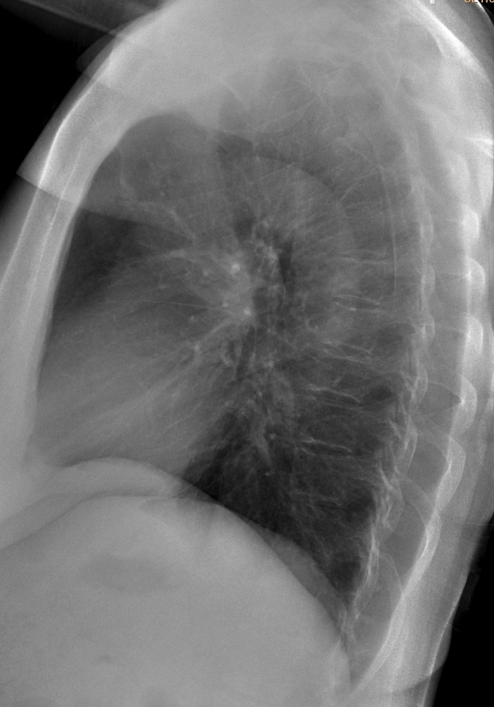
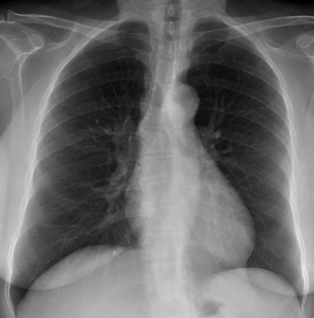
Muppet is in Chicago at the RSNA meeting. While looking for Miss Piggy, he found time to discover the following case: 65-year-old female asymptomatic.
Do you think the chest is normal? If not, then where is the abnormality?
Click here for the answer to Case 44
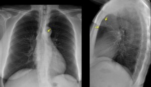
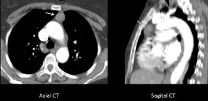
Findings: PA chest shows a mass in the left upper mediastinum (arrow), clearly visible in the anterior clear space in the lateral view (arrow). Enhanced axial and sagittal CT confirms the presence of a rounded soft-tissue mass in the anterior/superior mediastinum.
Final diagnosis: thymoma.
Remember that it is not unusual to find mediastinal masses in asymptomatic patients. Do not forget that small masses may not be obvious and have to be discovered. In the present case, findings are apparent if one looks for them.
Teaching point: always look at the anterior clear space in the lateral view of the chest. It can offer useful information as in this case and in case 27.


Thoracic kyphoscoliosis?Broncholithiasis.Marked small pleura interlobaris dex.Cardiomegaly.Arteriosclerosis aortae.Low position of the fornix of the stomach-may be because of a process under the left diaphragm….
No, il torace non è normale: esiste una opacità a livello mediastinico, al di sopra della carena tracheale a sx che impronta il bronco principale di sx.”While , mister Caceres looking Miss Piggy”, qui a Bari scopriremo cosa è!!!A presto.
subtle opacity in anterior segment RUL with air bronchogram (may be ground-glass at CT). In case this is not an acute condition, bronchoalveolar carcinoma should be ruled out.
There is a dislocation of the paraoesophageal line on the left.
Looks normal to me, bar the minor scoliosis.
It ain’t normal. Look carefully
The only thing I noted was prominent pulmonary vasculature…but in clinical practice it is quite probable that I would have reported this as normal!
I was also seeing a subtle nodular opacity just lateral to the left hilar point – which I am attributing to a superimposition.
I still blame it on Ms. Piggy!!
Perchè è stata proposta per “prima” la L.L. del torace? : è a livello della finestra aortico-polmonare la chiave di lettura? Ho 2 idee al proposito: è chiaro comunque che la TC fa la diagnosi.Il professore ha trovato Miss Piggy ?
Perché la L.L del torace? Because nobody look at it and can be very helpful.
Found Miss P. and had a wonderful time.
Abnormal. There is an opacity on left side of trachea near biforcation, (as Dr genchi says) in the area of aorto-pulmonary window and an irregular contour (lobulated) of azigos-esofageal stripe. So it should be an adenpathy.
This my first comment, however, I’m a big fan to Caceres’ corner as well as Dr. Pepe’s casebook.
Frontal: there is a small rounded density overlying the ascending aorta.
Lateral: barely seen mass in the anterior clear space (anterior mediastinum), so my impression is thymoma and better to be evaluated by CT.
For a first comment it is quite accurate. Congratulations.
Abnormal.
Although there is a scoliosis, the right edge of the mediastinum seems empty. On the left there are two abnormal structures over the aortic knob and the aorto-pulmonary window.
The description is rather inexact but I think it could be a persistent left superior vena cava.
We are expectant about Muppet and Miss Piggy.
Muppet is a gentleman and would not comment about a lady.
Would suggest that you look at previous blog cases. Answer may be there.
Thank you.
There is an anterior mediastinum mass. Tymoma?
See? It is not difficult if we know were to look. And the anterior clear space if one place to investigate, always.
I first considered the subtle retrosternal opacity as pulmonary. Now it seems it should be located in the anterior mediastinum, and the opacity noted by Bari, superimposed to the aortic knob must be a focal widening of the anterior mediastinal line.
La LL del torace non è perfetta perche, leggermente ruotata , per cui si vedono archi costali. Comunque l’opacità in paratracheale sx , potrebbe avere sede retrosternale ed allora la diagnosi più probabile, ricordando la legge dele 4 T per il mediastino anteriore, potrebbe essere un timoma asintomatico
The anterior compartment of the mediastinum is not homogenously radiolucent. Could this be a thymoma or thymolipoma?
Thymolipomas are usually bigger
Combination of PA and lateral Xray shows an anterior mediastinal mass + In the lateral Xray there is a linear structure from the left hilum to the mass.
This is probably a vessel (feeding vessel).
Although not in a typical location, this should be a extrapulmonary sequestration, with the feeding vessel arising propably from the left pulmonary artery.
(correction: extralobar sequestration)
Muppet is a simple fellow. Your diagnosis is highly sophisticated
I used to work in a University hospital. That’s why I like fancy diagnoses and sometimes forget the KISS rule of Muppet!
Good. See how well you fare with the next case.