Muppet wishes to present the case of a 75-year-old woman with bilateral mastectomies for carcinoma 10 and 7 years previously. Chest radiographs and CT are shown.
1. Pleural metastases
2. Mesothelioma
3. Pleural TB
4. None of the above
Answer case 51
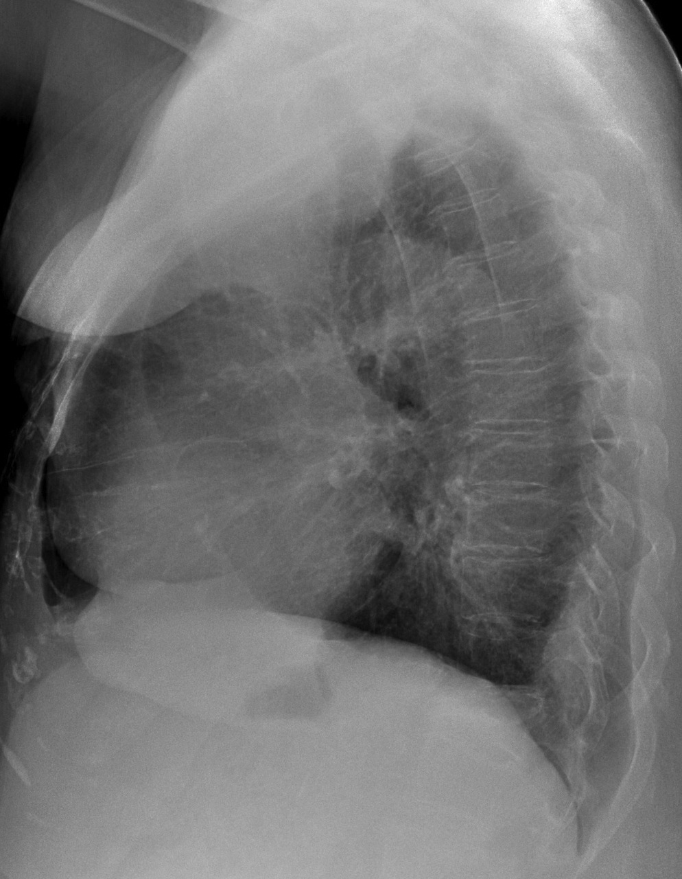
Findings: PA chest shows pleural thickening along the left chest wall (arrows, with blunting of the costophrenic angle and apical thickening. There is also an apical extrapulmonary lesion (red arrow). Lateral film suggests a second lesion (red arrow).
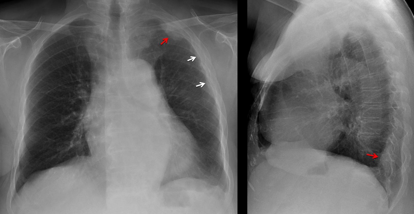
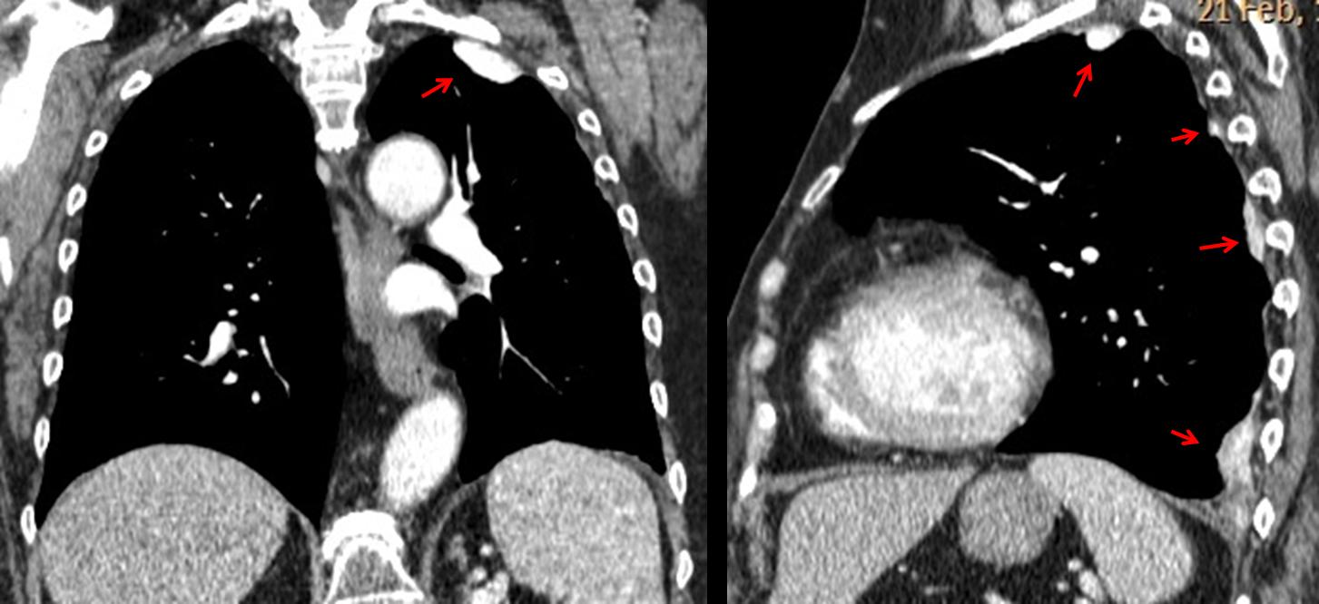
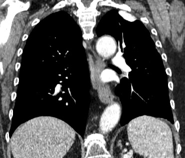
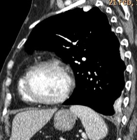
There is collapse of D-12. Sagittal and coronal CT show several high-density pleural-based lesions (arrows).
The differential diagnosis includes, among others, asbestos plaques, amyloid, pleural TB and calcified metastases after treatment. In an oncologic patient, another consideration should be talc pleurodesis. This patient had such a procedure in 2008 for recurrent pleural effusion. Appearance of chest two years later was unchanged.
Final diagnosis: talc pleurodesis
Congratulations to Dr. Genchi, who was the first to suggest the diagnosis
Teaching point: any particular finding (in the chest or otherwise) may have a iatrogenic origin. Don’t forget to ask if any procedure has been performed.


None of above
None of the above.
4. None of the above.
none of the above
Can you be more specific?
L’emitorace sx è debolmente rx-opaco rispetto al controlaterale; inoltre in AP si nota una ipertraspareza ” a semiluna” in corrispodenza dell’arco aortico, mentre in LL si osserva una tenue opacità in sede retrosternale, delimitata posteriormente da linea pleurica.In AP sembra raddoppiata la silhouette dell’aorta discendente.Sulla TC in coronale,massa di tessuto solido che occupa il bronco lobare superiore sx.Penso allora ad atelettasia del lobo polmonare superiore sx da metastasi mammaria.
Sorry, CT is incomplete. There was no bronchial mass. I am concerned about the pleural lesions
Seems like pleural thicking on left apex as well as lower left lobe……. Not sure:-)
Pleural TB or could be an asbestosis associated pleural plaques
la signora ha avuto versamenti pluerici recidivanti a sx? come sono stati trattati?(pleurodesi chimica-Talcaggio?)
I think Genchi is right. This lady must have had pleural metastasis years ago (T12 is colapsed) and treated with talc pleurodesis. As a result there is an apical talc pleural nodule and fibrous hyperenhancing posterior pleural nodules.
Pleural nodules are enhancing a lot. This called my attention. Generally metastasis do not have this pattern. I think this patient can have another thing (but I have no idea).
Good! So you will learn something from this case.
Focal pleural thickening on left apex and posterior costofrenic sulcus. The chest X-ray also shows some left diffuse pleural,thickening (not shown on CT). We should have an unenhancet CT to evaluate the degree of enhancement.
I think there was a left pleural effusion at some point.
The diagnosis might be fibrin bodies or fibrous calcifiing pseudotumor
It could be a pleural fibrous tumor. They are often pedunculated and in many cases also depend on the visceral pleura. Furthermore they are not related to exposure to asbestos and rarely calcify.
May present intrapulmonary feigning injury and normally have a homogeneous enhancement in administering contrast CT study when they are small.
How many times have you seen multiple pleural fibrous tumors?
ever.
I take it back
never
I take it back.
(Sorry for my bad English)
Grazie galactico!!!!
This pleural lesion show intense contrast enhancement , it take nearly same contrast as arteries at the arterial phase , this finding not consistent with methotheliomas , metastasis or pleural T.B . I think it is another pathology and Iam thinking of another pathology likely vascular lesions because of that intense enhancement