Dear Friends,
My colleague Jordi Andreu, who is almost as mischievous as the Muppet, presents the following case: a 45-year-old woman with pain in the chest.
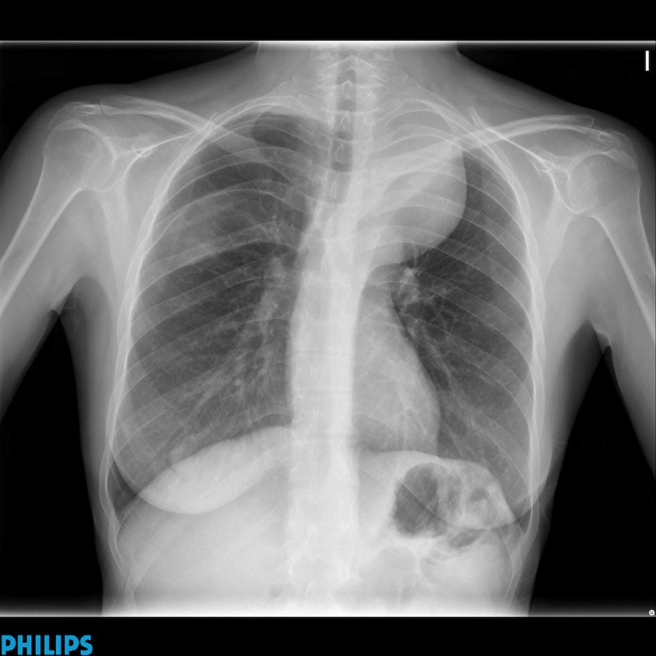
45-year-old woman, PA chest
Diagnosis:
1. Neurogenic tumour
2. Thymoma
3. Fibrous tumour of pleura
4. None of the above
Click here for the answer to case #54
This case shows interesting semiologic findings. There is an obvious left mediastinal mass, which extends to the lung apex, placing the mass in the posterior mediastinum. It cannot be a thymoma because the upper mediastinum ends at the level of the clavicles. A goiter can be excluded because the trachea is not displaced. In addition, there is remodelling of the right fifth rib (arrows) and ribbon-like appearance of the right and left third ribs (arrows). All these findings point to neurogenic tumours in a patient with neurofibromatosis.
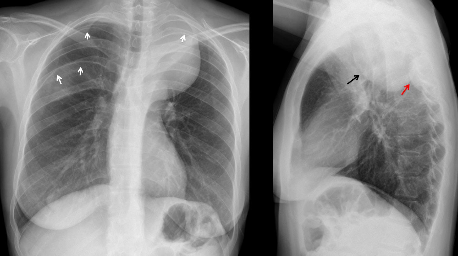
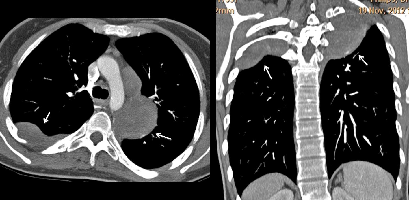
The lateral view shows the large mass (arrow) and a smaller one (red arrow), which is responsible for the remodelling of the right fifth rib. CT confirms the presence of both masses (arrows).
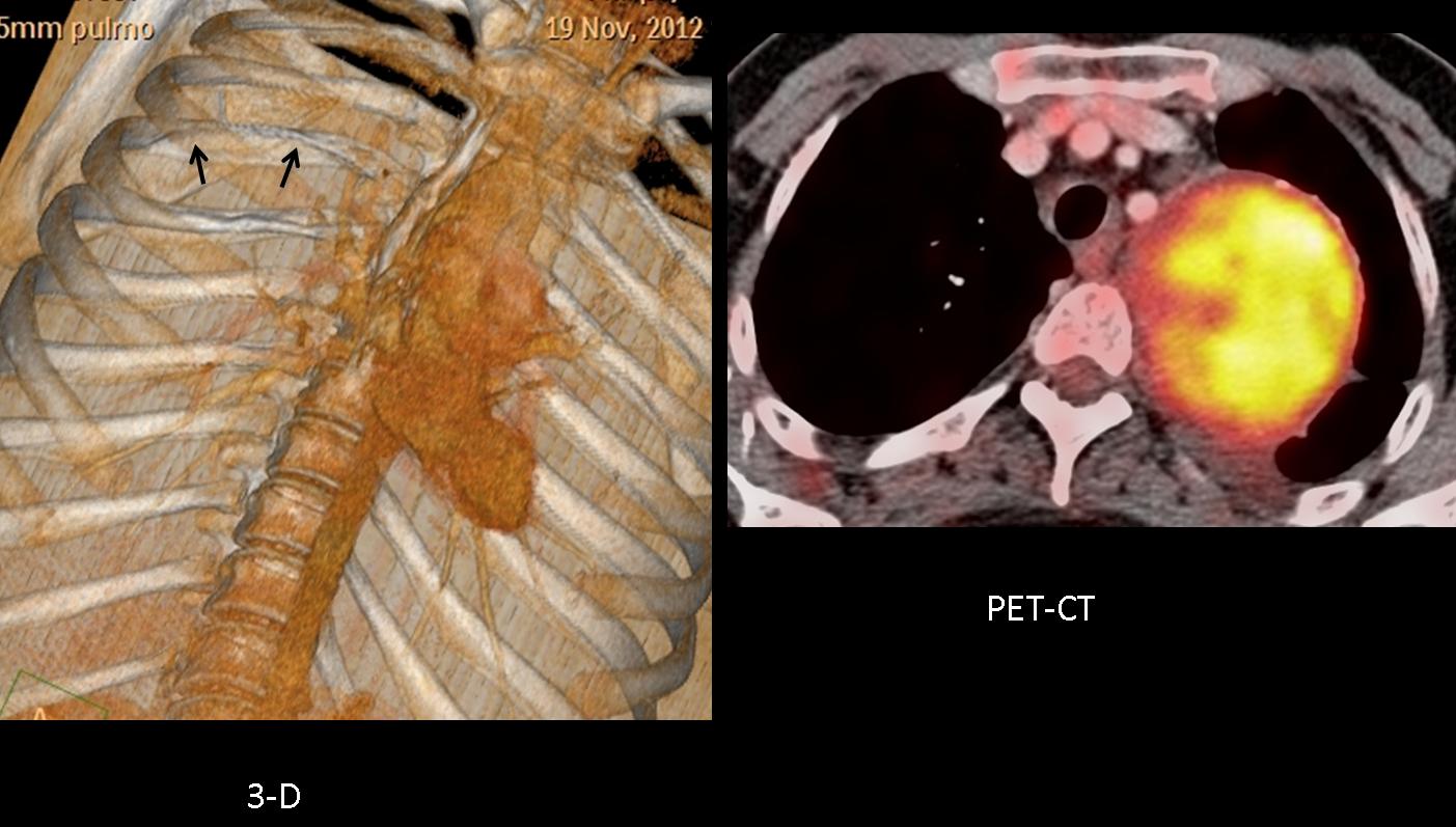
Remodelling of the right fifth rib is better demonstrated in the 3D reconstruction (arrows). PET-CT shows high uptake of the left mass.
Final diagnosis: neurofibromatosis type I, with malignant degeneration of the big left chest tumour.
Teaching point: posterior mediastinal masses accompanied by rib notching and ribbon-like ribs are highly suspicious of neurofibromatosis.


I think it is a fibrous tumour of pleura.
I think it is a neurogenic tumour (large neurofibroma) in type 1 of neufibromatosis.
I also think it’s rather a neurogenic tumour. A large fibrous tumor of pleura would have more acute angles. Though the age group(40-60 decade)is suitable. And the mass extends above the clavicle, so it can’t be in the anterior mediastinum.
It appears like a left mediastinal tumor, probably a neurogenic tumor. There are also fibrotic findings and a possible area of increased opacity at the RUL. Hazy border of the inferior part of the posterior arch of the right 5th rib. I conclude to large neurogenic tumor (neurofibroma), with a possible past medical history of excised intercostal neurofibroma. Neurofibromatosis
Esclerosis de los bordes inferioroes de varias de las costillas en el lado derecho. Quién sabe, a lo mejor tiene algo que ver con una coartación de la aorta.
neurogenic tumor
Schwannoma tipo A di Antony. Perche’? Una opacità di massa omogenea( non calcificazioni mixoidi) ed a contorni netti, posizionata nel mediastino posteriore( allargamento della linea paraspinale).Esiti di rimaneggiamento chirurgico della 5 e6 costa di dx, con velatura della parete toracica( per esiti fibrotico-cicatriziali), con sclerosi lungo il bordo costale inferiore della 5( decorso dei nervi intercostali).Il neurofibroma ha margini più irregolari e si configura negli altri segni della NF tipo 1 che non sono menzionati.Opzione per il Ganglioneuroma.
neurogenic tumor.. ribbon ribs on the right.. NF 1
The left upper zone mass has sharp and obtuse margins, hence extrapulmonary. the aortic knuckle is seen through the mass and the mass is seen extending above the clavicles, hence it is a posterior mediastinal mass. Also the bilateral 2nd and 3rd ribs are slender with inferior notching of the posterior aspect of the right 3rd rib. The inferior margin of posterior aspect of the right 3rd rib is also indistinct. The unifying diagnosis for posterior mediastinal mass, slender ribs and inferior rib notching, is Neurofibromatosis type I, hence option 1 is the answer.
Excellent discussion. Congratulations. You qualify for a free gift during ECR.
Thank you, I am following your corner for last 6 months. your teaching methods have radically changed the way I approach chest imaging. In fact, this blog helped me a lot during my preparation for FRCR 2b exams.
Domanda: come si fa diagnosi di NF tipo 1 se non sono presenti gli altri segni della Rechkinghausen( all’Rx-torace) . Es. a livello toracico una scoliosi e-o una displasia fibrosa scheletrica?PS : il Bari malissimo anche in serie B:rischio di retrocessione.
Sorry, I am now in Sicily and cannot find documentation. But I was taught that posterior mediastinal tumors plus ribbon-like ribs were highly suspicious of neurofibromatosis
Stimatissimo professore, non sono d’accordo.La mia 2 specializzaione è la Pediatria ( la 3 è la medicina sportiva).La diagnosi di NF, tipo 1 ( ci sono 7 forme di NF, di cui il tipo 1 è certamente la più frequente)è “clinica” e si basa sulla presenza di almeno due dei sette aspetti clinici della stessa.La presenza di neurofibroni, cutanei, sottocutanei e-o dei nervi profondi , in numero almeno superiore a due è uno solo dei sette aspetti clinici. le alterazioni ossee a dx possono essere dovute agli esiti di rimaneggiamento post-chirurgico alla rimozione di un neurofibroma cutaneo.Le alterazioni ossee nella Reckinglhausen sono altre: a livello toracico è prevista solo la scoliosi.Con stima e l’affetto di sempre.
thin ribs on the right!!! Probably neurogenic tumor.