
Dear Friends,
Muppet is getting old and feeble and, after more than one year of showing cases, is exhausted. He has decided to stop searching for exclusive cases and has decided to show only cases seen within the last two weeks. Hopes they are still worth presenting and will earn your approval. The first case, seen on April 15, is the PA chest radiograph of a 57-year-old woman with previous breast cancer, removed. What do you think of the anterior right fifth rib?
Diagnosis:
1. Metastases
2. Fibrous dysplasia
3. Paget’s disease
4. None of the above
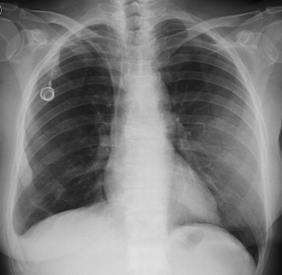
57-year-old woman, PA chest
Click here for the answer to case #64
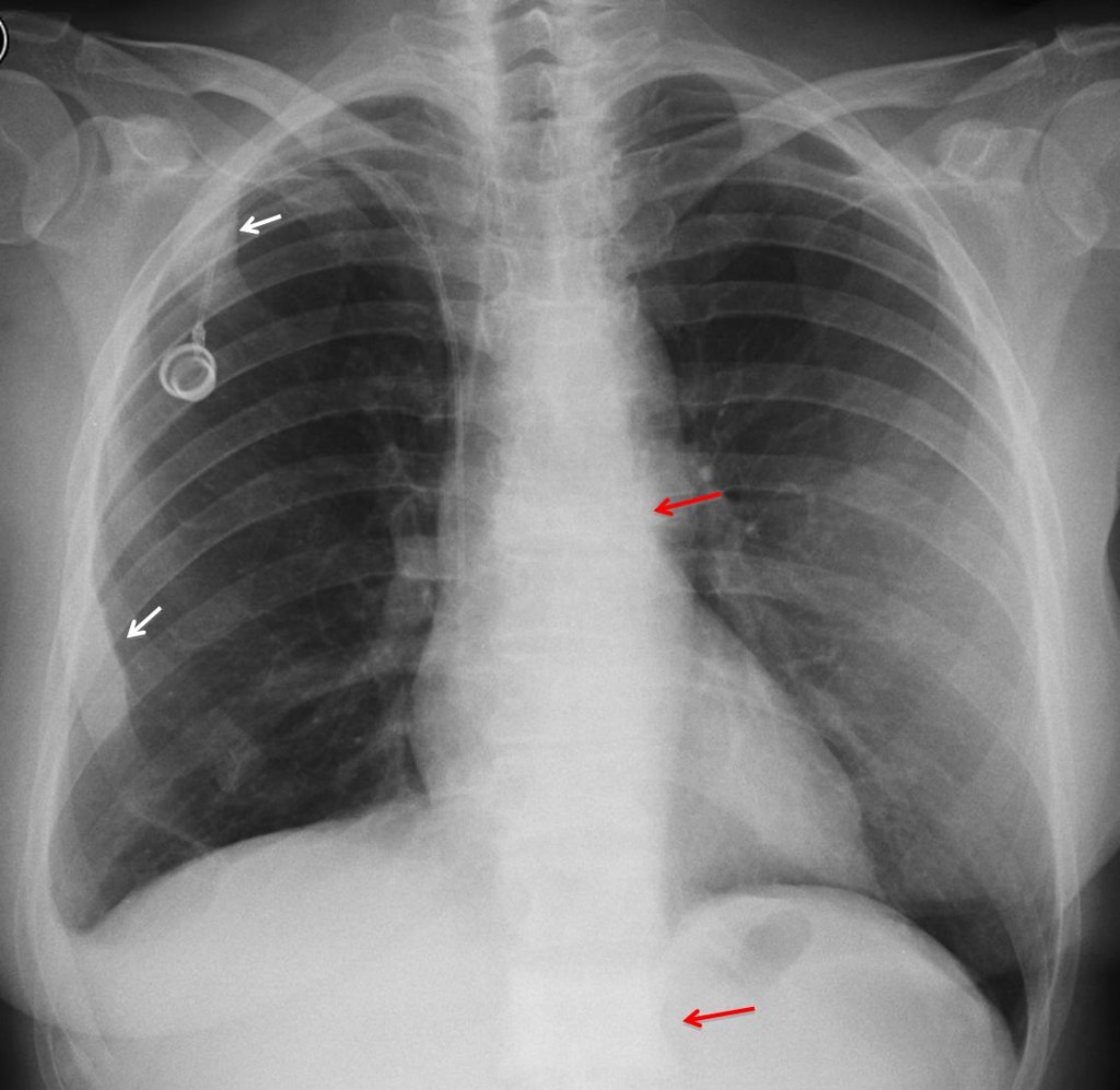
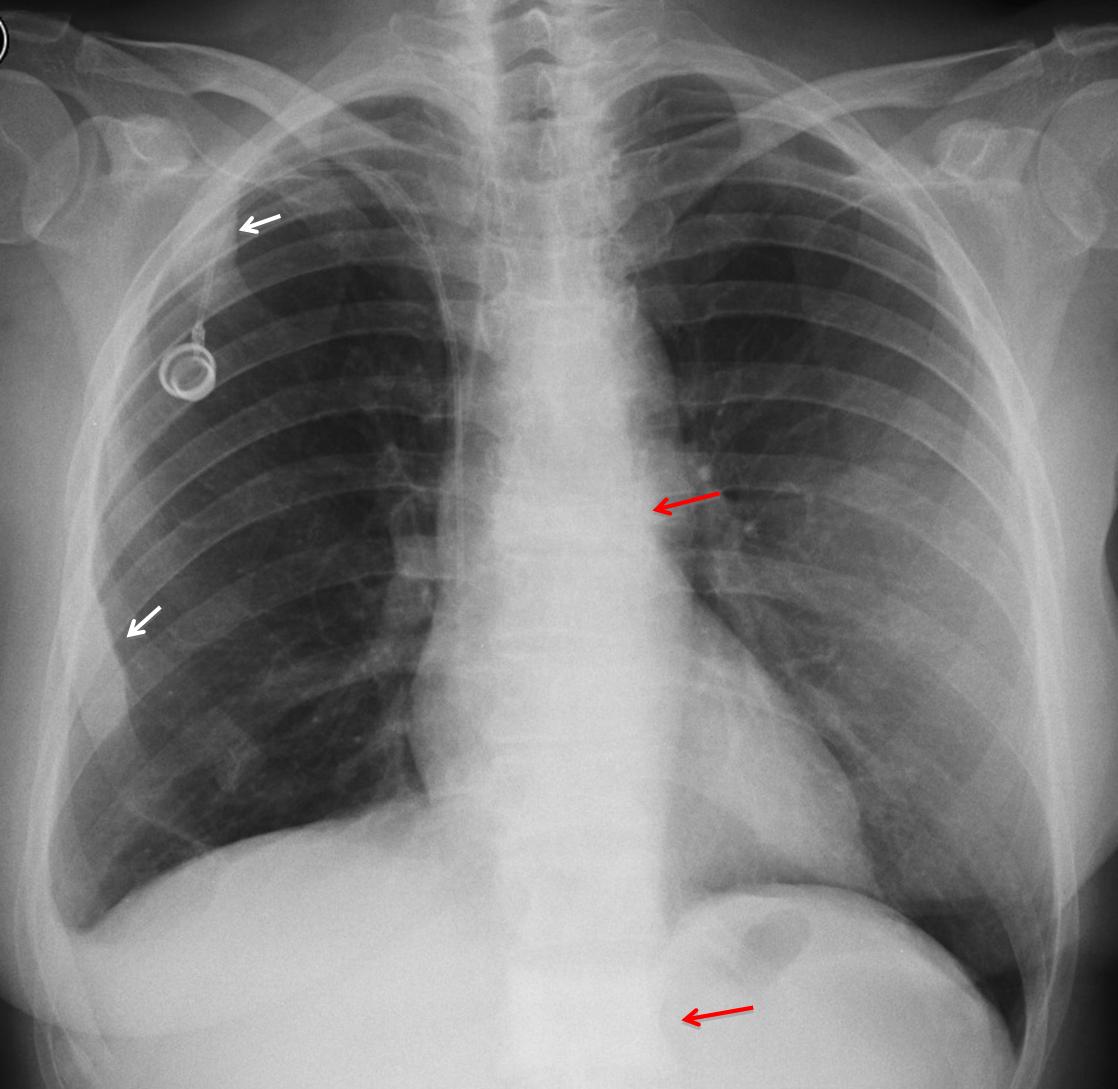
Findings: the PA radiograph shows increased density of the anterior aspect of the right 2nd and 6th ribs (arrows). There is collapse of T-8 and sclerosis of L-1 (
red arrows). These findings are highly suspicious of osteoblastic metastases, especially in a patient with breast carcinoma.
Sagittal and coronal CT confirms the widespread sclerotic changes of the spine, affecting the right scapula as well.
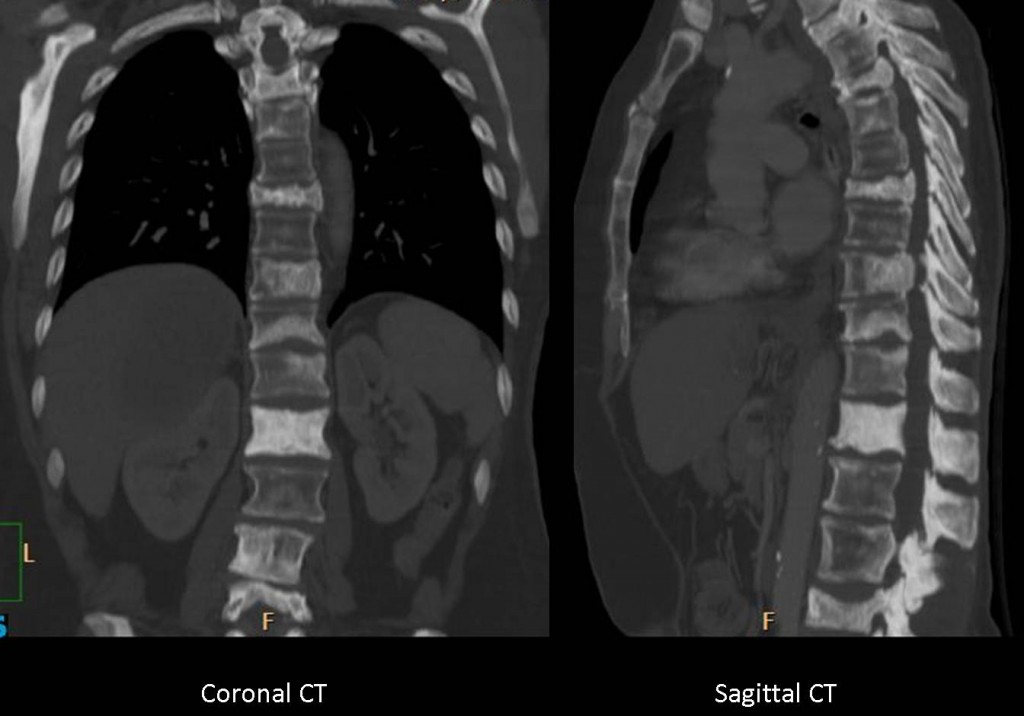
Final diagnosis: osteoblastic metastases from breast carcinoma
Congratulations to Katerina, who was the first to answer and gave the right diagnosis.
Teaching point: always consider a common manifestation of a common disease. In this particular case, Paget’s disease and fibrous dysplasia are a distant second choice


The lesion in the anterior right 6th rib (if I am counting right) is an homogeneously sclerotic expansile lesion with sharp borders, no cortical destruction and no associated soft-tissue mass.
The differential diagnosis for a solitary sclerotic expansile lesion in the rib is osteoblastic metastasis, Paget disease, osteoid osteoma, osteoblastoma and healed brown tumor (fibrous dysplasia has a ground-glass appearance and therefore it is excluded).
But, I think that this lesion is not solitary because there are 2 more homogeneously sclerotic expansile lesions in the anterior right 2nd rib, in the anterior right 8th rib and maybe a sclerotic appearance of L1.
So, taking into account the history of the patient, I choose
1. Metastases (osteoblastic)
PS. The diagnosis would be easier if we could compare this x/ray with previous ones
It’s the sixth rib. I stand corrected.
Loss of right costophrenic angle ,Pleural effusion?
brest cancer left or right?she had radiation therapy?
I think we have increase in opacity of a vertebral body (L1)
osteosclerotic (Ivory) Vertebra and (T7)maybe colapse of (T7).
Sclerotic expansile lesion of the anterior right 6th rib (i think katerina is right) and anterior 2th right rib.
Osteitis deformans is more common polyostotic but frequent sites of involvement are spine,pelvis,skull and proximal long bones.There is relatively common over the age of 80 > men
blastic phase is on late state (inactive)
We have informations of a bone scintigraphy?
She likes the hats?:)
more information profesor
Bone scintigraphy will be shown in due time.
Nobody wears a hat nowadays (except Muppet and myself)
Sclerotic lesions of discrete segments of II, VI e VIII right ribs, bone enlargement and cortical thickening: Paget (blastic).
But i’m a shameful optimist.
I ll go for polyostptic Paget and ignore the right costophrenic angle blunting, which is rather due to old pleuritis or surgery.
Given the clinical scenario is the first choice osteoblastic metastases.
A good option, if we compare with previous studies, would be the possibility of changes post-radiotherapy and / or chemotherapy prior lytic metastases.
In conclusion osteoblastic metastasis or metastasis aftertreatment lytic changes of breast cancer.
she had implants in both breasts? or she operate on the left?
Patient has an implant in the left breast
La domanda del professore è precisa: cosa pensare dell’arco anteriore della 6 costa dx. Essa tuttavia deve essere valutata nel contesto clinico( CR mammario) e dell’imaging “accessorio”( lesione osteosclerotica della 2 costa dx , “vertebra “d’avorio” di L1 e deformazione sclerotica del soma di D8.Valutando con le” precedenti” immagini, se prima non c’era nulla l’ipotesi prima è per metastasi osteoblastiche.Ma…l’arco anteriore della 5 costa oltre che addensato è aumentato di volume e questo depone contro la metastasi che porta a diminuzione di volume…..
osteoblastic metastases
AVN following radiotherapy, or sclerotic metastasis.
poliostotic Paget in diferent phases