After spending a long weekend with Miss Piggy, Muppet is at peace with the world and has chosen an easy case: 41-year-old male with cough, mild fever and left chest pain.
1. Pneumonia
2. Infected bronchiectasis
3. Pulmonary infarction
4. None of the above
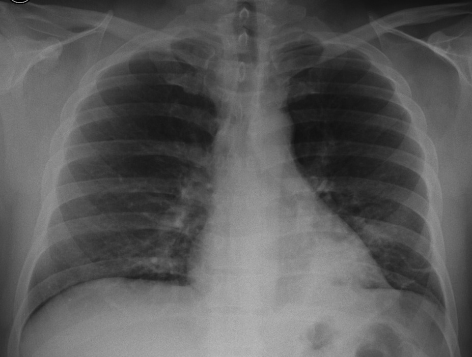
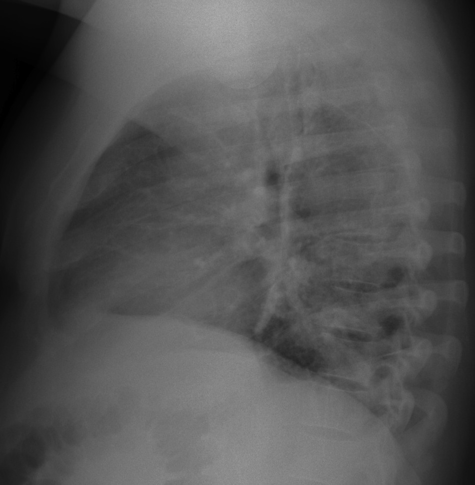
Chest radiographs taken in 2009 show a peripheral opacity in the RLL (arrows). There is marked downward displacement of the left hilum (
red arrow), indicating volume loss.

Fig. 1
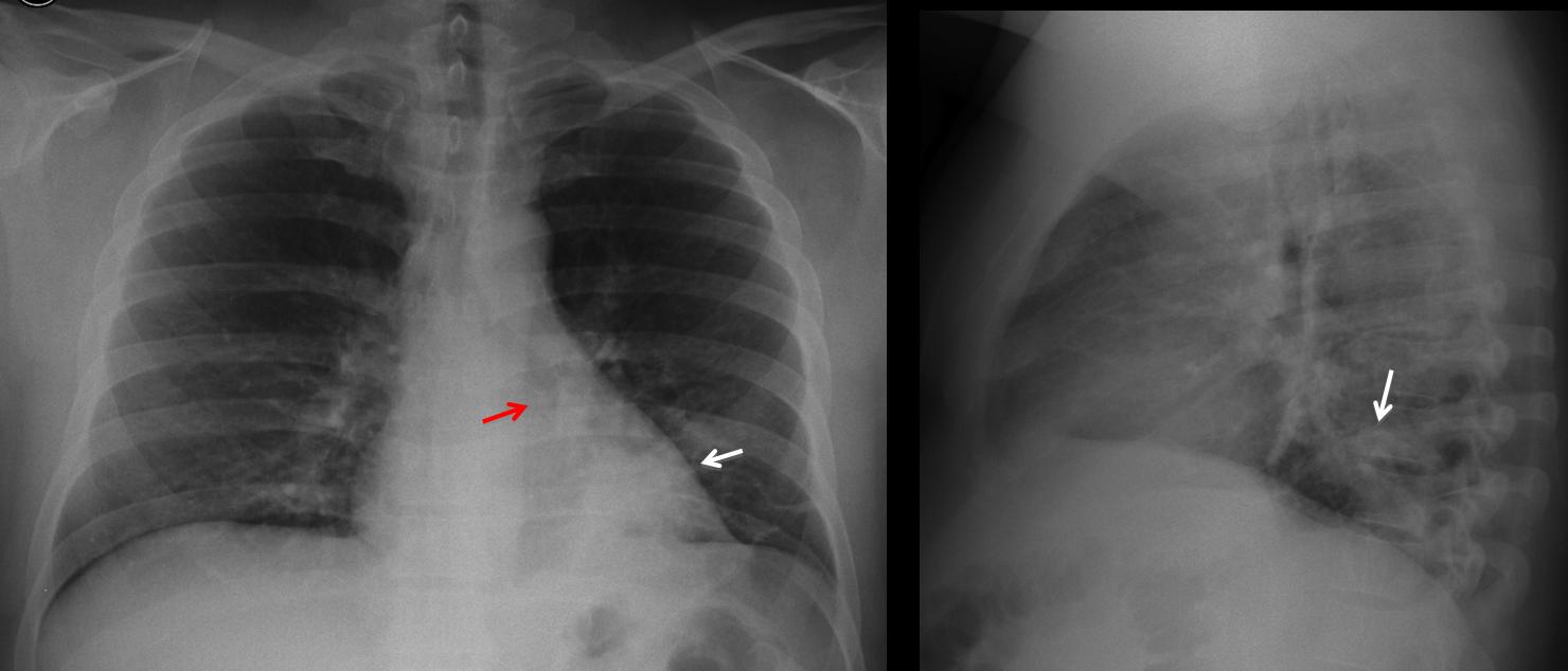
Hilar displacement was overlooked and the patient did not have new radiographs until February 2013, showing an obvious LLL collapse (Fig 2a, arrows). CT demonstrates an endobronchial lesion (2b, arrow), with fat in the center (2c, arrow). There is marked collapse of LLL (b, c, red arrows). A lobectomy was done.
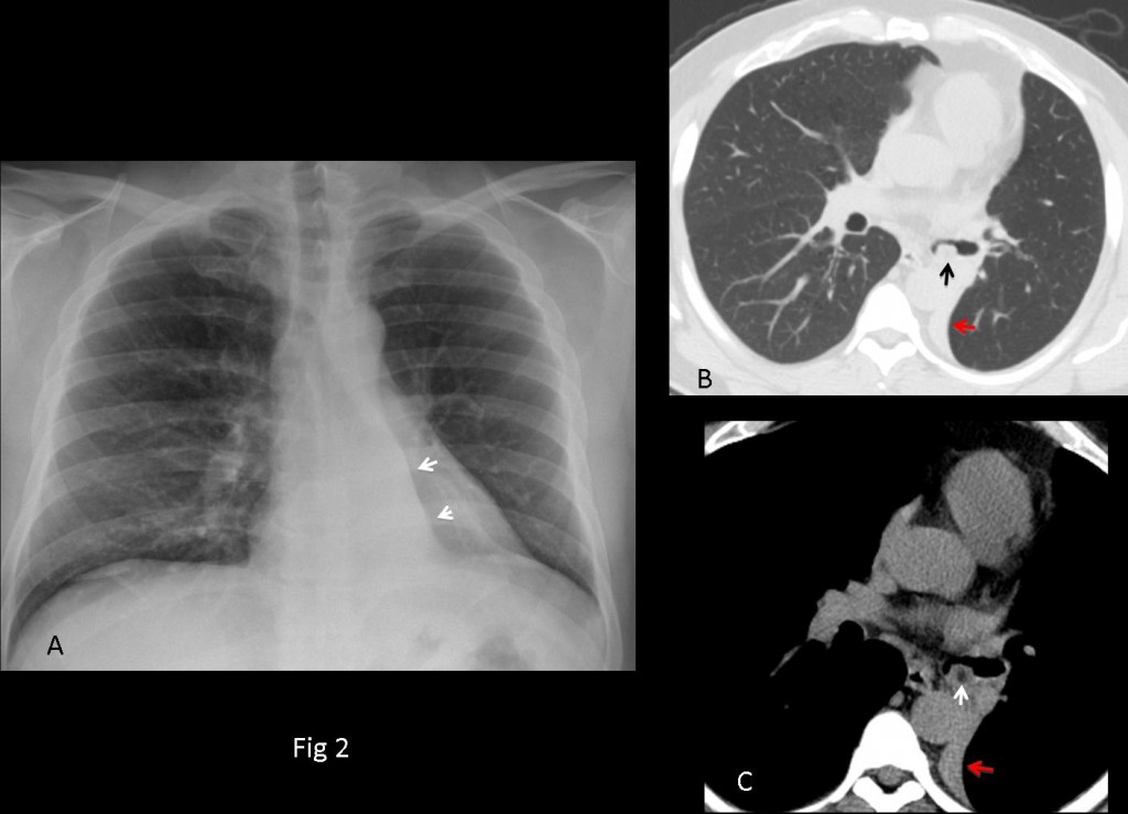
Fig. 2
Final diagnosis: endobronchial hamartoma
Congratulations to Dr. Mapia, who first mentioned the downward displacement of hilum.
Teaching point: don’t forget to look at the hila. In this patient, the diagnosis was delayed four years and it cost him a lobectomy.


Pneumonia: lower lobe consolidation behind the heart, silhoutte sign.
.Окръглено пневмонично огнище в областта на лява белодробна основа, с ангажиране на диафрагмалната плевра-Pleuropneumonia sin.Incl.-Bronchitis chr.Bronchiectasiae bill..
Google translates as: Rounded pneumonic outbreak in the left lung base, with involvement of diaphragmatic pleura. I take you vote for number 1.
Πύκνωση στο οπίσθιο κάτω αριστερό πνευμονικό πεδίο συμβατή με πνευμονία (pneumonia) η βρογχογενές καρκίνωμα ( Ca)
Google traslation: Thickening at the rear lower left lung field consistent with pneumonia (pneumonia) the bronchial carcinoma (Ca)
Left lower lobe consolidation-mass with left hilum shift downwards. Lung carcinoma is a possibility.
2. Infected bronchiectasis “Gloved finger” sign?
2. Infected bronchiectasis
Opacità a “dito di guanto” a carico dei segmenti basali laterali e posteriori del LLL , da” mucoid-impact” , per brochiectasie cilindriche, frammiste ad immagini bronchiolo-alveolari( non vi è iperinsufflazione del territorio polmonare circostante:non atresia bronchiale).Regola KISS. Il Bari e’ Salvo dalla serie C.
KISS rule is useful when you take all the findings into account. Did you look at the hilum?
Congratulations about Bari!
Chest x ray frontal nd ael view .
An irregular branchng opacity is seen at left lower lobe associAted with non homogenous air dpace acification silhouetting left medial hemidiaphragm , with increased density over the lower thoracic spine , no pleural. Effusion , so it is sugestive of inflammatory process , infected bronchiectasis .
Ripartiamo: le opacità a “dito di guanto” rappresenterebbero sempre “mucoid-impact” mentre l’addensamento disomogeneo circostante potrebbe andare per una polmonite “ostruttiva”, il che spiegherebbe la posizione anomala dell’ilo come da processo produttivo endobronchiale.
That is much better. Answer tomorrow.