Dr. Pepe’s Diploma Casebook: Case 107 – To err is human: how to avoid slipping up (Chapter 6) – SOLVED!

Dear Friends,
To conclude the section “To err is human” I am presenting PA radiographs of a 57-year-old hairdresser with interstitial lung disease, who is on the waiting list for lung transplant. What do you see?
Check the images below, leave your thoughts in the comments section and come back on Friday for the answer.
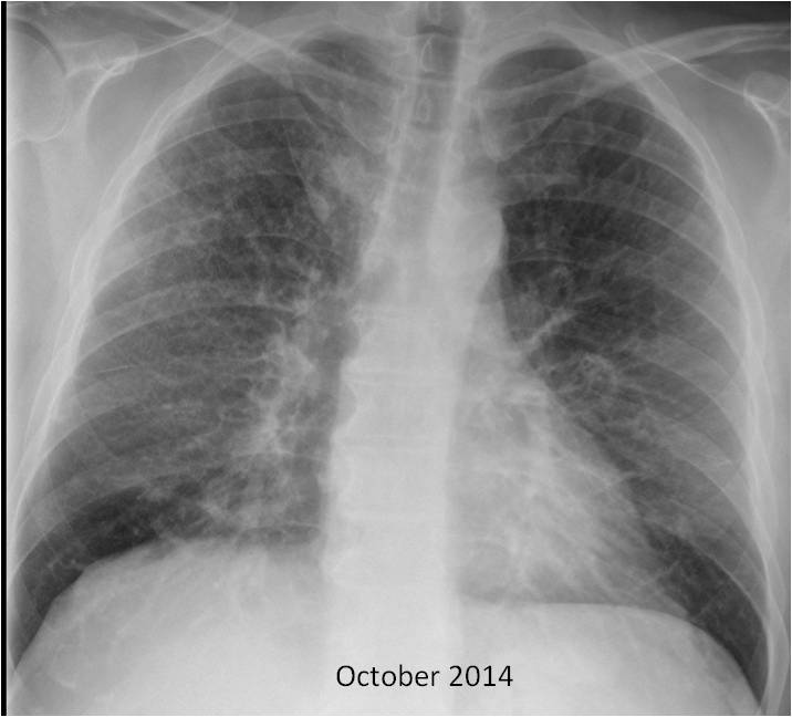
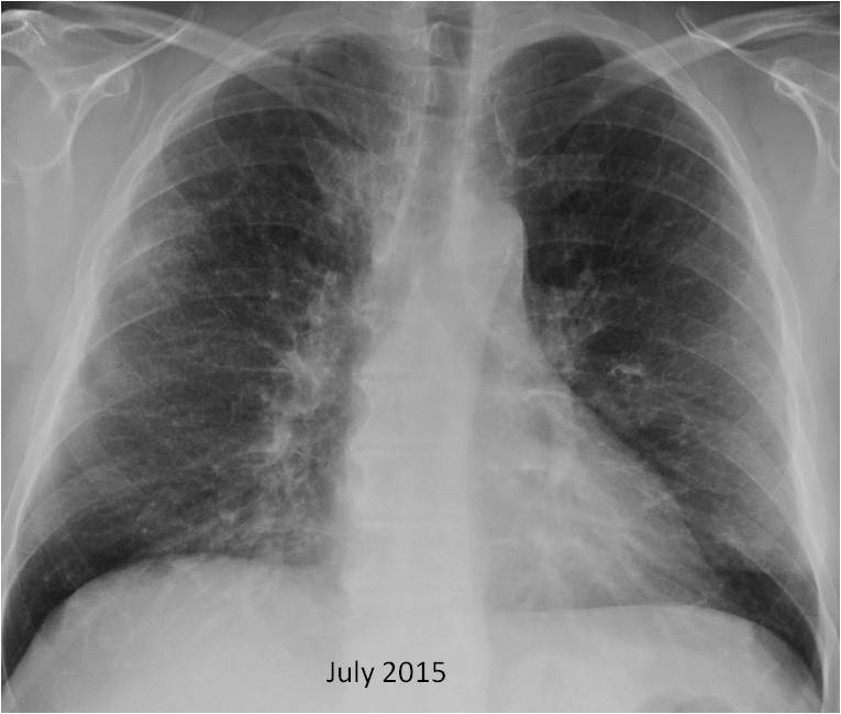
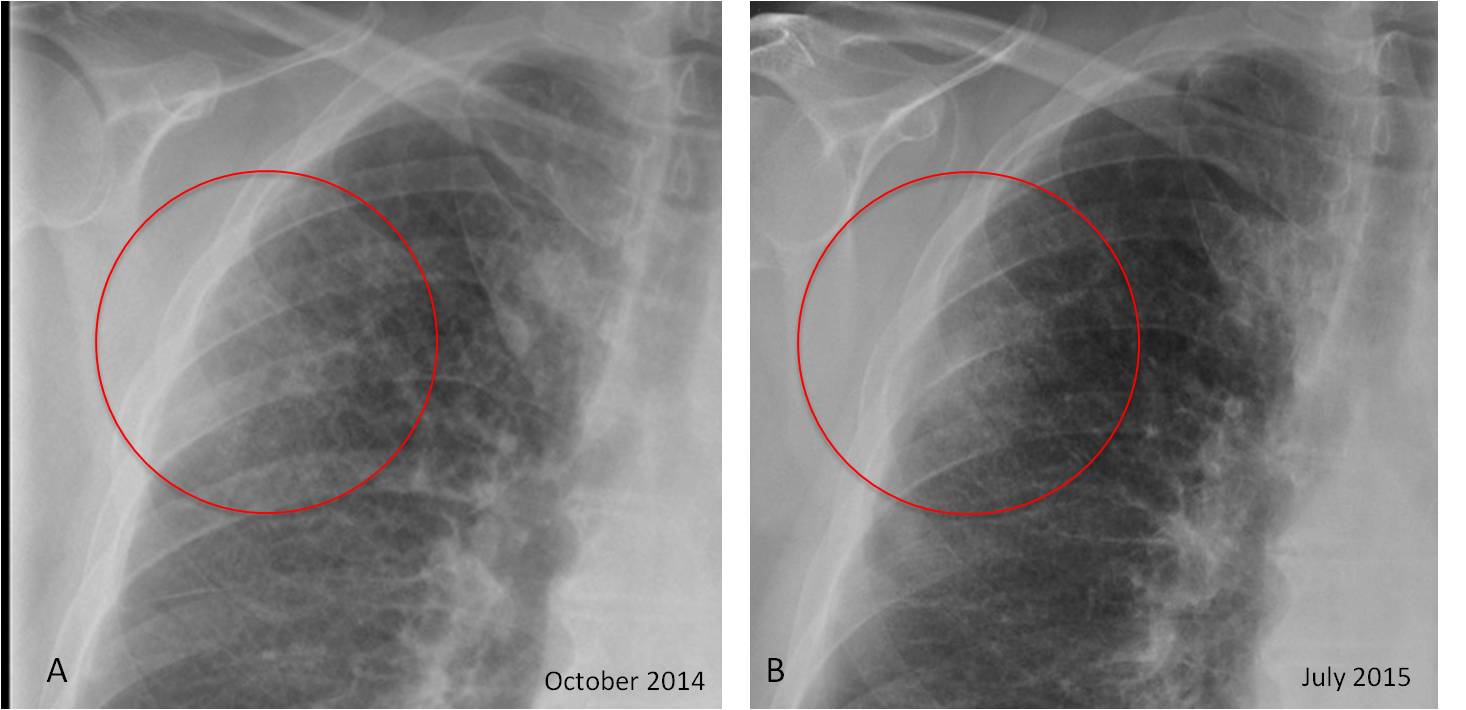
Findings: chest radiographs show diffuse pulmonary infiltrates, consistent with the clinical information. In addition, there is an area of increased opacity at the periphery of the right lung (A, circle) which has become more evident in the follow-up study (B, circle).
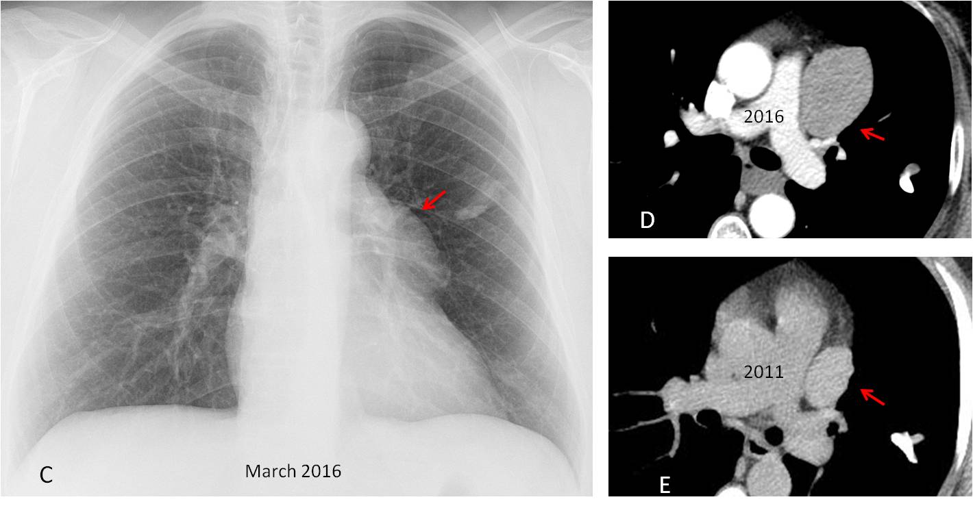
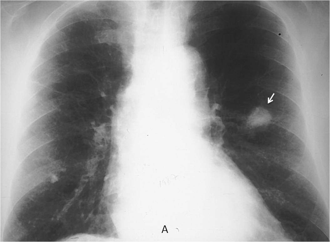
The opacity was obvious by the next month (C, arrow) and was reported by the radiologist. However, the surgeon was not aware of this development and scheduled the patient for surgery. Fortunately, the opacity was noticed in the preoperative radiograph (D, arrow) and the intervention was cancelled. Needle biopsy demonstrated a carcinoma, which responded to chemotherapy (E, arrow).
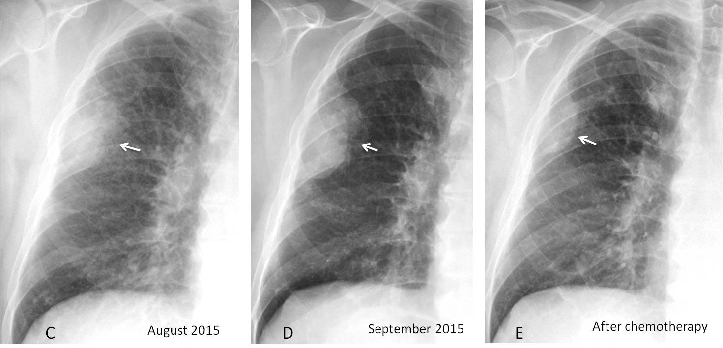
Final diagnosis: fast-growing carcinoma in a patient with interstitial disease
In this final chapter I want to comment about errors in communication. These occur at all levels in imaging departments (secretaries, clerks, technicians), but the most important one is when a correctly interpreted and reported abnormality fails to reach the referring physician, causing a delay in the diagnosis that may be detrimental to the patient. Today’s leading case is a good example of this type of error.
In one study, 38% of communication errors had an impact on patient care. Most of them were due to faulty communication between the radiologist and referring physician. This is why the American College of Radiology states that urgent findings and other findings that may impact the patient’s health require direct communication with the referring physician.
To emphasise the importance of correct communication, I’ll show several representative cases.
Case 1 depicts a preoperative chest radiograph for bariatric surgery in a 51-year-old woman. A tubular opacity was detected in the left lung (A, white arrow). The convex border of the left mediastinal contour (A, red arrow) was overlooked. Unenhanced CT shows that the pulmonary opacity represents a calcified mucus impaction (B, white arrow) and additionally discovers a solid anterior mediastinal mass (B, red arrow), reported as a possible thymoma.

Case 1
Five years later, a second preoperative radiograph shows that the mediastinal mass has increased in size (C, red arrow), and this is confirmed with enhanced CT (D and E, red arrows). The mucus impaction remains unchanged.
This is a good example of communication error. The surgeon probably did not read the report in 2011 and the radiologist failed to alert him about the finding. On surgery in 2016, a benign thymoma was found.
Case 1
Case 2: preoperative chest films for urethral repair in a 26-year-old man. A large opacity of uncertain etiology is seen in the lower right hemithorax (A and B, white arrows). There is a loop of bowel in the right upper quadrant (A, red arrow), which could be due to Chilaiditi syndrome or elevation of the hepatic flexure, secondary to diaphragmatic hernia.
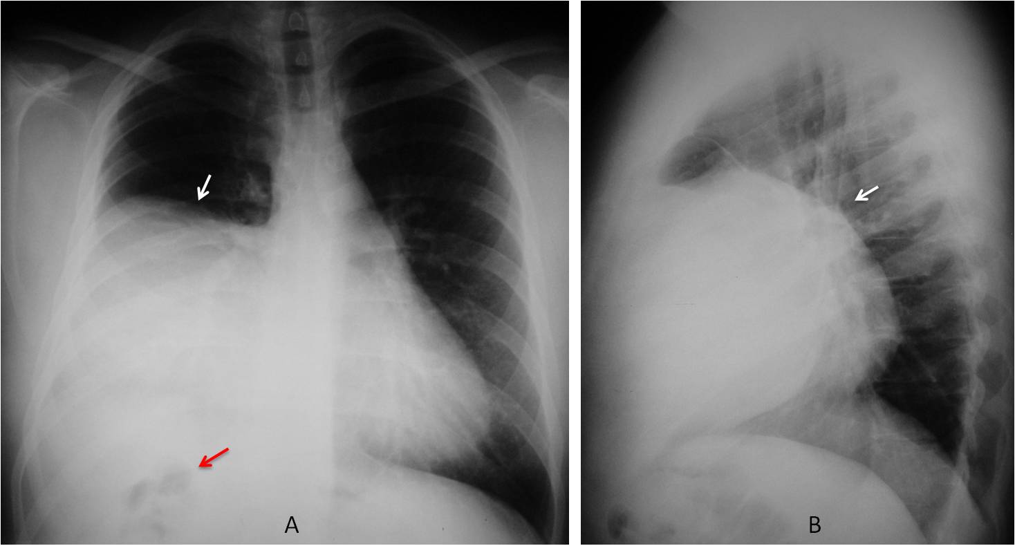
Case 2
Review of the clinical history showed that the urethral stenosis was secondary to rupture of the urethra and bladder neck in a motorcycle accident four years earlier. Chest films taken two years before the present one showed apparent elevation of the right hemidiaphragm (C and D, arrows). The changes over time and background of trauma suggested diaphragmatic hernia as a good possibility.
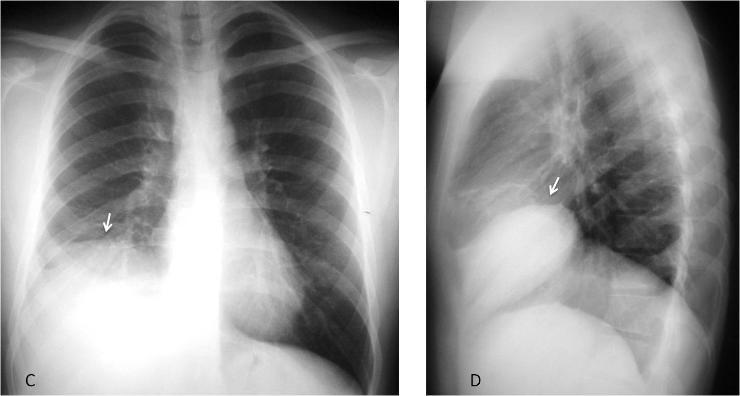
Case 2
The patient was recalled and coronal and sagittal unenhanced CT showed a rent in the diaphragm (E and F, arrows) with herniation of the liver. In this case the referring physician was responsible for the communication failure. The radiologist had called repeatedly to report the unexpected findings, but surgery did not ensue. The patient finally underwent surgical treatment 17 months after the CT and confirmation of traumatic hernia.
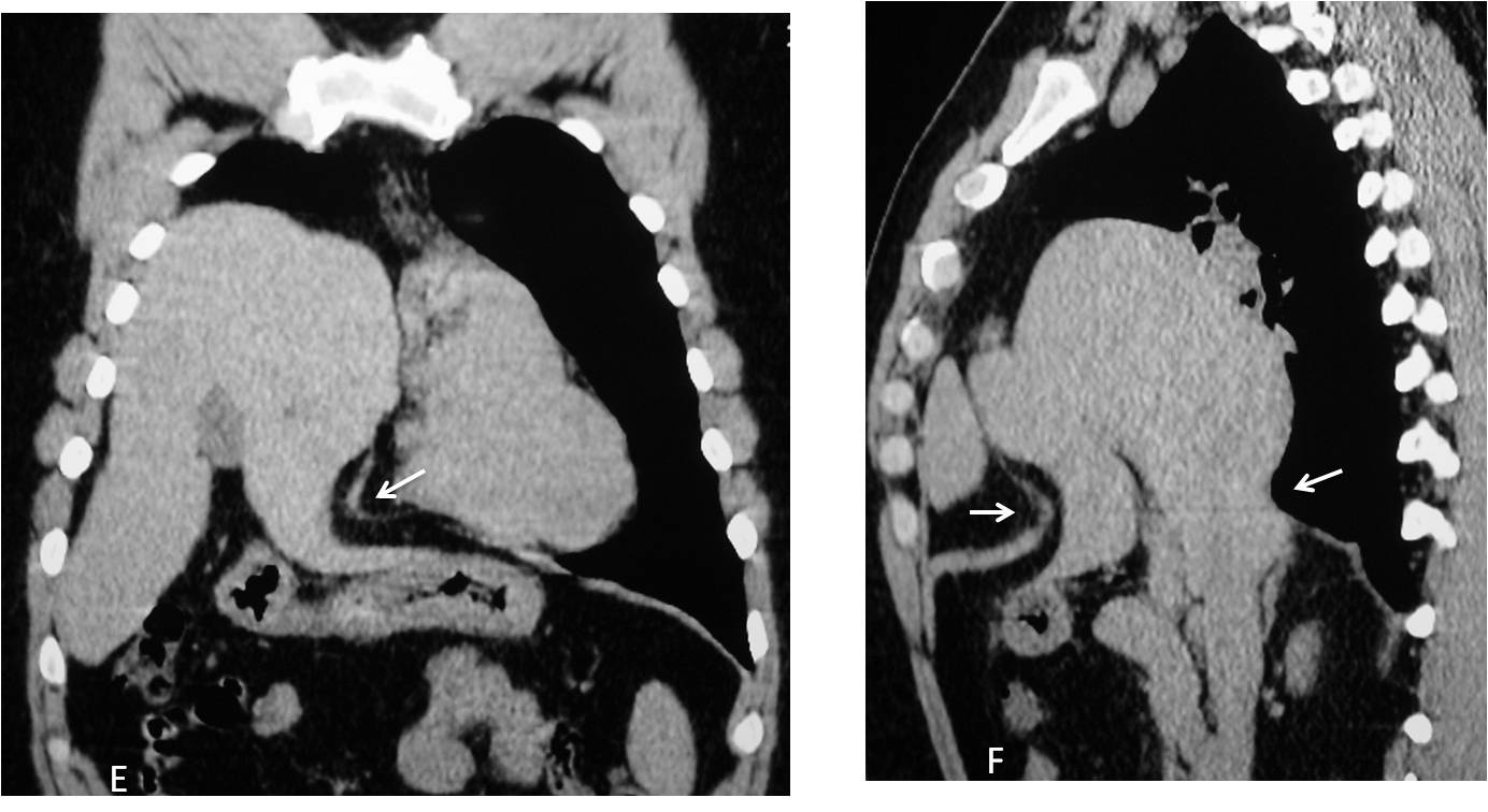
Case 2
Case 3 is a vintage example. This radiograph, taken in 1983, belonged to a 72-year-old man who was having a femoral bypass. The radiologist mentioned the nodule in the left lung (A, arrow), but the surgeon failed to read the report or look at the radiograph. After surgery, the nodule was noticed and needle biopsy confirmed adenocarcinoma. No abnormal lymph nodes were found at chest surgery. The patient lived another eight years and died of unrelated causes. He was my future father-in-law.
Case 3
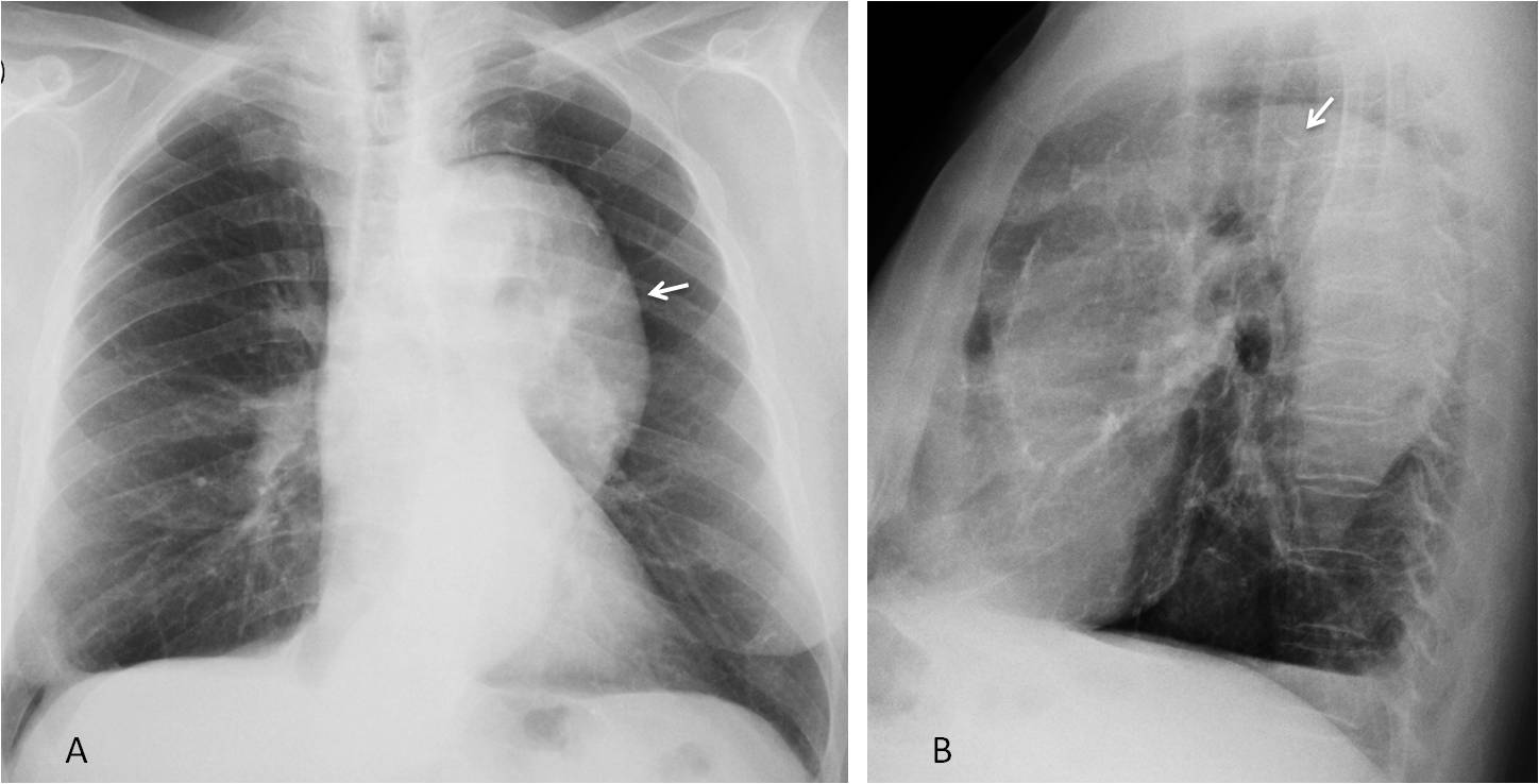
To avoid communication errors, we should directly notify the referring physician and keep a record of the cases, to insist if no action is taken. In Case 4 we see the typical finding of type B aortic dissection, a very prominent descending aorta (A, arrow), with a possible displaced atheromatous plaque in the lateral view (B, arrow). This case was seen in January 2017 and the referring physician was duly contacted. So far, no CT has been carried out. It may be time for a kind reminder.
Case 4
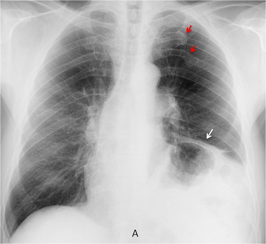
Case 5: preoperative chest radiograph shows marked elevation of the left hemidiaphragm, with an irregular contour (A, white arrow). In addition, there are old fractures of the left 4th and 5th ribs (A, red arrows). This combination of findings, as in case 2, suggests traumatic diaphragmatic hernia, which should be ruled out or confirmed with CT. The patient was seen in May 2016, but so far, no CT has been done.
Case 5

Follow Dr. Pepe’s advice:
1. Errors in communication can have a major impact on patient management.
2. Most of them occur between the radiologist and referring physician.
3. They are corrected by directly contacting the physician when a significant abnormality is found and keeping a log to remind them if no further action is taken.


hello professor.
In this xray there are reticular opacities in bilateral lungs right > left likely fibrotic changes.additionally, cystic and tubular lucencies in the right lung predominantly in parahilar region representing bronchiectatic changes. Lower zones are relatively spared. Follow up xray after an year shows reduction in the opaciites – ? post steroid. however, there is increased/ persistent irregular opacity in the right middle zone along with the pleural thickening along the right lateral thoracic wall In a patient of ild ,I would be concerned to rule out a malignancy. hence would advise a ct scan. Of incidental note is the azygous fissure.
Good morning,
Peribroncovascular thickening, more visible at paracardiac margins because of intersticial pathology.
Acigos fisure is present.
Hypertrophic of the first rib articulation.
Like Ren, I think there is a sutile right pleura thikening not visible in previous xray.
Dear Dr. Pepe
Additionaly to all the fibrotic changes I think there’s a mass in the right middle pole that grew twice over the year.
Regards
Dear Professor,
Maybe posttherapeutic reduction in reticular opacities in both lungs.
I´m also concerned of a subtle hazy density in the right lateral lung field corresponding to a pleural mass, DD: coarse lung malignant lesion. Underlying ribs seem to be intact.
….Stimatissimo Prof. …la patologia è a carico del lobo accessorio v: azygos…..la scissura era ben visibile nella radiografia 2014….non più visibile nel 2015, per probabile retrazione atelettasica del lobo, da processo produttivo…..simpaticissimo prof. ti avevo avvertito che Dybala sarebbe stato il vostro incubo….per il mio Bari, sempre più figure inguardabili….
You were right and I was wrong
Thanks to all five brave people that participated. Kudos to Ren, who was the first to do the right diagnosis.
More information tomorrow