Dr. Pepe’s Diploma Casebook: Case 13 – SOLVED!
Dear friends,
This week’s case is a sixty-year-old male with progressive exertional dyspnea.
Most likely diagnoses:
1. Arrhythmogenic right ventricular dysplasia
2. Tricuspid valve endocarditis
3. Ebstein anomaly
4. Giant aneurysm of the right atrium
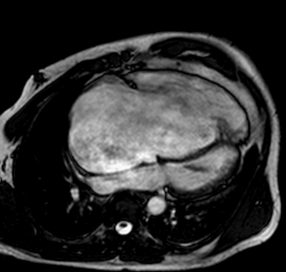
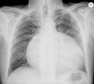
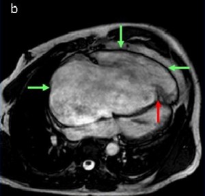
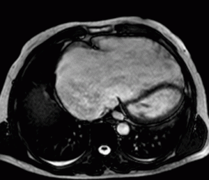
Findings: (a) Chest radiograph depicts marked cardiomegaly with normal pulmonary vessels. (b) Steady-state free precession cine-MRI in four-chamber view shows apical displacement of the septal tricuspid valve leaflet (red arrow), atrialisation of the right ventricle, and considerable dilatation of the right atrium and right ventricle (green arrows).
Inferior ectopic displacement of the septal tricuspid valve leaflets is the hallmark of Ebstein anomaly. The right atrium and the base of the right ventricle (atrialisation of the right ventricle) are enlarged.
Right ventricular enlargement is one finding of arrhythmogenic right ventricular dysplasia/cardiomyopathy, but the tricuspid valve is in a normal position.
Tricuspid valve stenosis can be associated with right atrial enlargement, but the right ventricle is normal.
Giant aneurysm of the right atrium, also known as idiopathic dilatation and congenital enlargement of the right atrium, is a rare anomaly characterised by massive right atrial dilatation, usually associated with tricuspid annular dilatation and tricuspid regurgitation. The right ventricle is normal.
Final diagnosis: Ebstein anomaly
Ebstein anomaly is characterised by varying degrees of dysplasia and displacement of the tricuspid valve leaflets into the right ventricle. The anterior leaflet is usually attached in the normal location, but is often large and dysplastic, and the distal leaflet is attached to portions of the right ventricular apex. The right ventricle is divided into an enlarged proximal portion and a more distal component with decreased pumping capacity. The effective tricuspid valve orifice is enlarged and inferiorly displaced into the right ventricle.
Ebstein anomaly is usually associated with right ventricular dysfunction and tricuspid regurgitation. More than 50% of patients have an atrial shunt, either a patent foramen ovale or a secundum atrial septal defect (ASD).
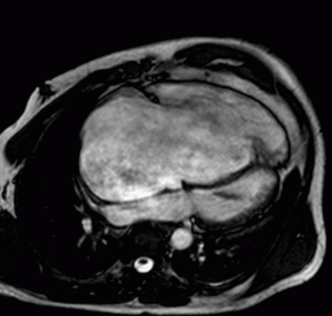
(Fig.5) Four-chamber axis and (Fig. 6) axial plane steady-state free precession cine-MRI images show displacement of the septal tricuspid valve leaflet towards the apex, atrialisation of the right ventricle, and considerable dilatation of the right atrium and right ventricle. Note the abnormal hypointense jet flow from the right ventricle to the right atrium during systole.
Ebstein anomaly and tricuspid atresia are both congenital abnormalities of the tricuspid valve. Imaging findings are characteristic and diagnostic.
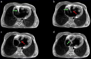
Twenty-year-old female with exertional dyspnea who underwent a Fontan operation for tricuspid atresia. (a-d) Axial T1-weighted MR images show atresia of the tricuspid valve with fat in the right atrioventricular groove (green arrow). Note hypoplasia of the right ventricle and a muscular interventricular septal defect (red arrow).
- Ebstein anomaly is characterised by varying degrees of dysplasia and displacement of the tricuspid valve leaflets into the right ventricle. The right ventricle is divided into an enlarged proximal portion and a more distal component with decreased pumping capacity.
- Enlargement of the right chambers is characteristic and is the reason for the abnormal appearance of the heart in the x-ray
- This causes of tricuspid regurgitation due to processes that directly affect the tricuspid valve, as occurs in rheumatic heart disease, infectious endocarditis, myocardial infarction, metastatic carcinoid syndrome, trauma, and Marfan syndrome.
Suggested reading:
Yalonetsky S, Tobler D, Greutmann M, et al. Cardiac magnetic resonance imaging and the assessment of Ebstein anomaly in adults. Am J Cardiol. 2011;107:767-73. PMID: 21247528
Case prepared by Rafaela Soler, MD




Giant aneurysm of the right atrium.
Dal radiogramma in AP avrei detto versamento pericardico:la RM , secondo il piano delle 4 cavità cardiache, chiarisce la estroflessione aneurismatica originante dall’atriop dx, con comprressione e disolocazione delle restanti cavità cardiache.
Wrong age group for Ebstein’s – such patients should present in the peri-natal period.
I cannot see any signs of endocarditis around the visible bit of the tricuspid valve.
We can barely make out the right ventricle (it being so small as compared to the right atrium)…let alone comment on it being dysplastic.
The right atrium is huge! It is compressing the left heart…with deviation of the atrial septum towards the left (indicating very high right atrial pressures). So I would consider the possibility of a congenital aneurismal right atrium!
Besides that, on the CXR, there is upper lobe venous diversion together with an interstitial infiltrate…quite suggestive of decompensated CHF.
I don’t that you can exclude Ebstein just with age alone
Epstein anomaly
Ebstein anomaly
Tricuspid valve insuffiency acquired [in 60 old male ](endocarditis?)with valvular plane dilated
there is also an increased marking vessels in lungs’s fields with congestive vascularity pattern.
Giant aneurysm of rt atrium .ebstein anomaly not possible in this age.
Dra. Soler (who prepared the case) answers: Ebstein may occur in adults. See the paper by Yalonetsky in the Am J Cardiol 107:767-773, 2011. In 31 patients range of age 21 to 68 years.
Thanks!
i would say Ebstein, had seen a case that was told to present at the age of 46.