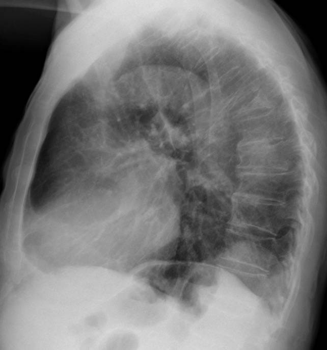
Showing chest radiographs of an 81-year-old male with multiple bone fractures after a car accident. There is a rounded well-defined opacity in the posterior costophrenic sulcus. What do think it is?
1. Carcinoma of the lung
2. Bochladek’s hernia
3. Diaphragmatic cyst
4. All of the above
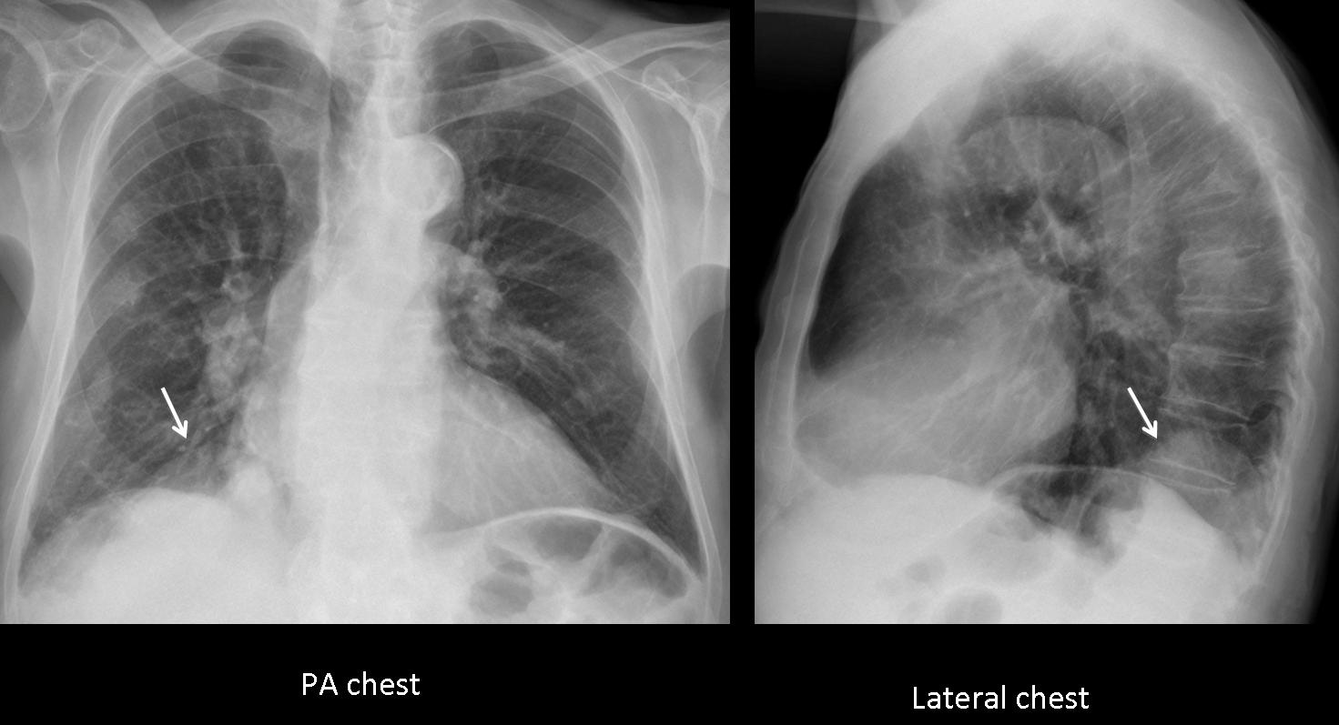
Fig. 1
Findings: aside from multiple rib fractures and a T-6 compression fracture, there is a bulge in the posterior aspect of the right hemidiaphragm (arrows).
The diaphragm is a frontier organ, and localised bulges in its contour may be due to alterations of the diaphragm itself (eventration), pleural or lung tumours, or a condition arising from below (hernia).
The most common lesion in this area is Bochdalek hernia, which is not infrequent in elderly people. However, a diaphragmatic cyst cannot be ruled out because conventional chest radiography cannot distinguish among the densities of fat (hernia), fluid (cyst), and soft tissue (tumour). A well-defined lung tumour cannot be excluded because the costophrenic space is narrow and the differential characteristics of intra- vs. extra-pulmonary lesions cannot be applied.
Therefore, the correct answer is: 4. All of the above
In this particular case, coronal and sagittal CT demonstrate a necrotic pulmonary mass occupying the costophrenic angle (arrows). Diagnosis: carcinoma of the lung, confirmed at surgery.
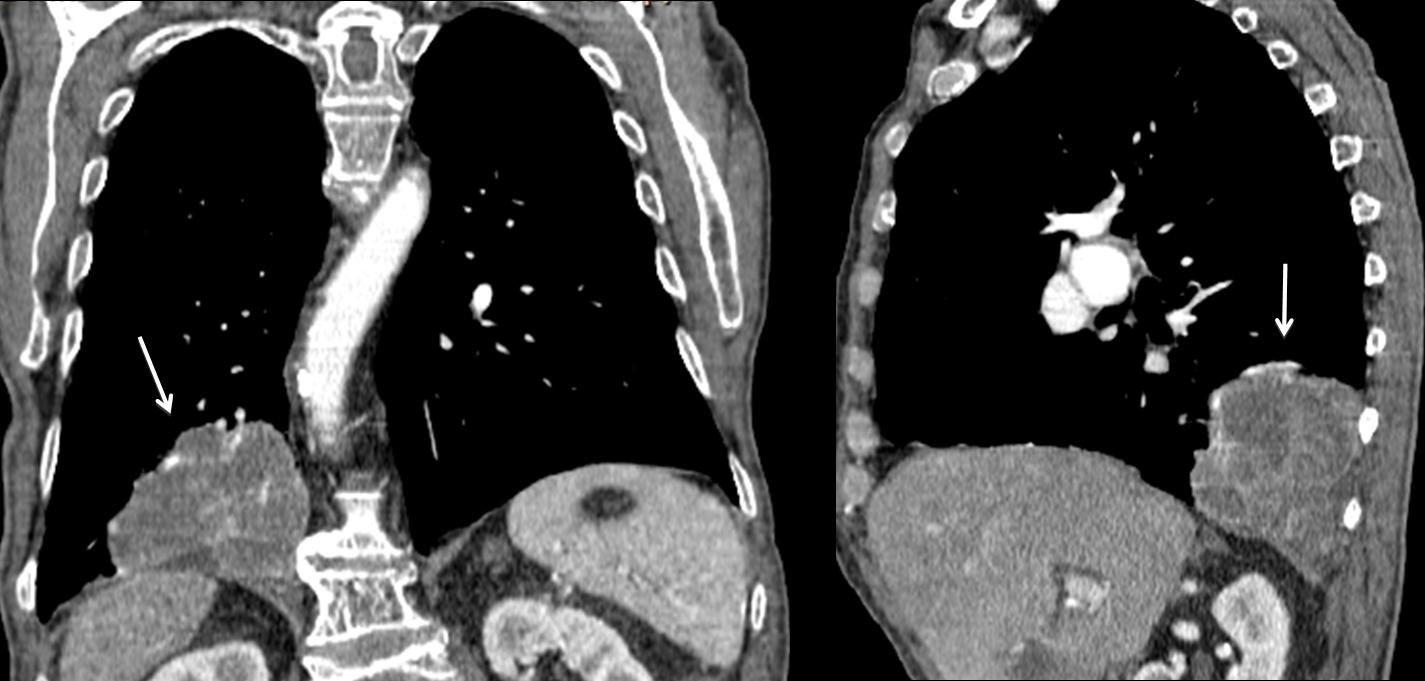
Fig. 2
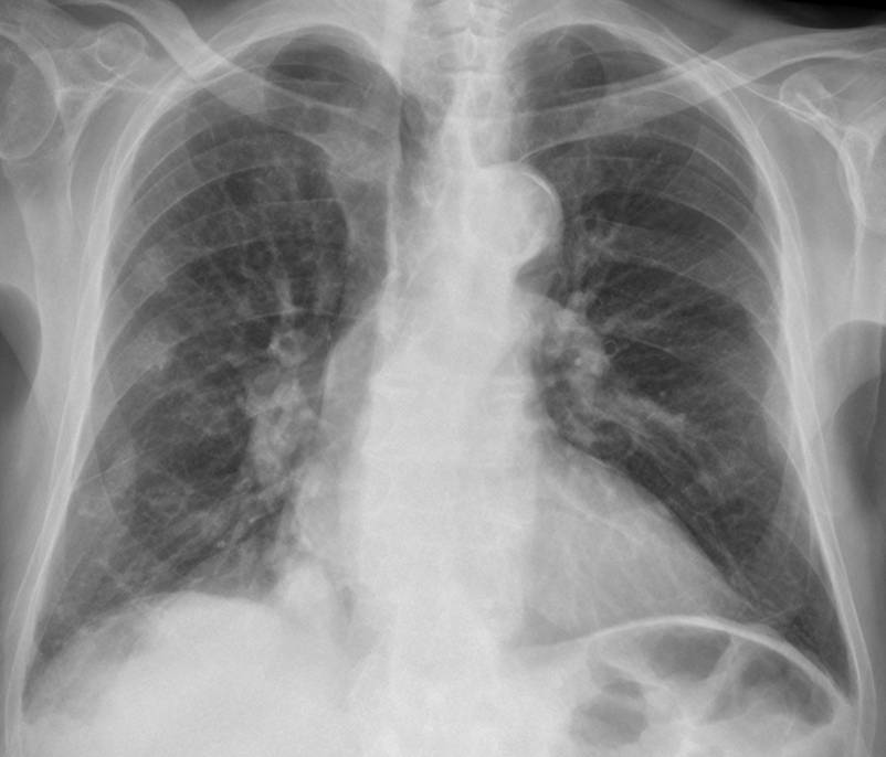
Occasionally, lung tumours may develop in the posterior costophrenic sulcus, as in the case below, which corresponds to an asymptomatic 56-year-old man. Chest radiographs show findings similar to the previous case, with a rounded opacity in the posterior sulcus (Fig. 3, arrows) which simulates diaphragmatic pathology.
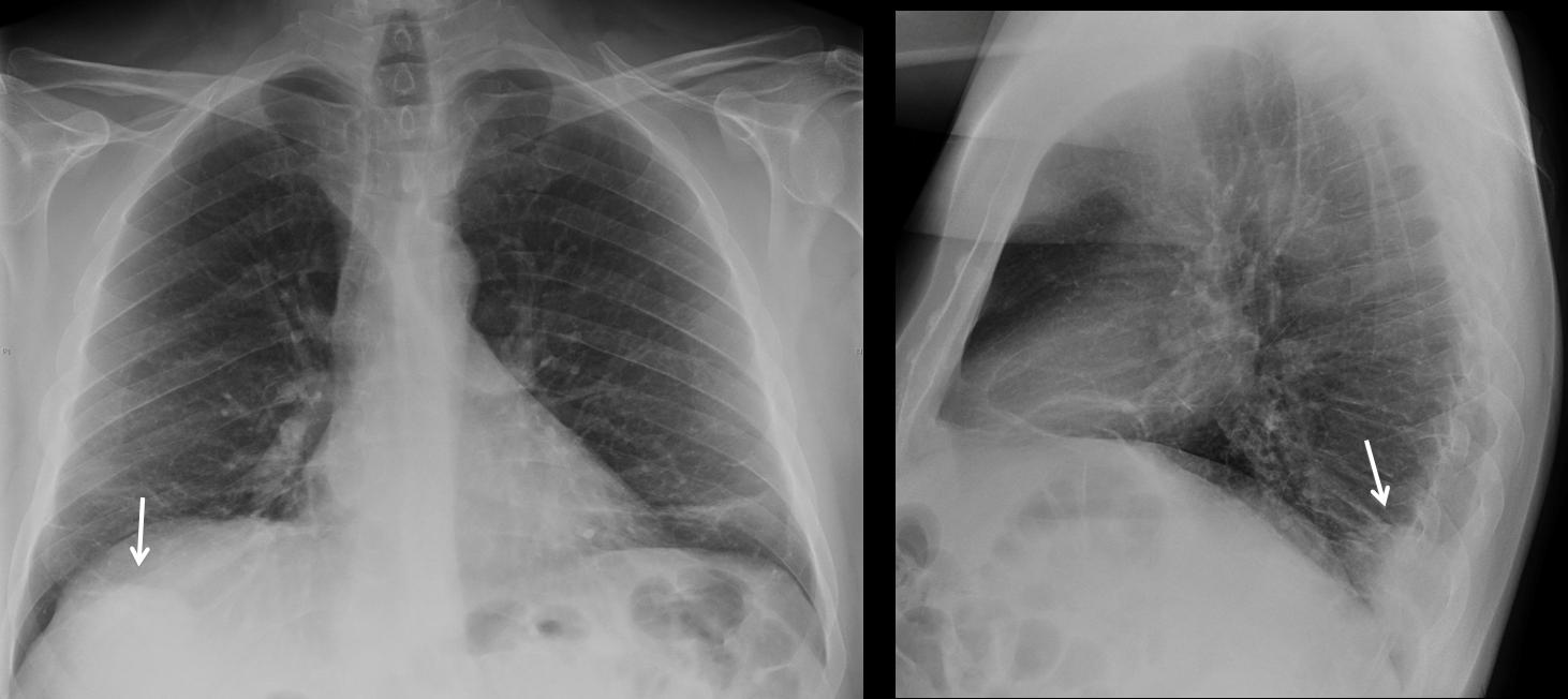
Fig. 3
Coronal and sagittal CT demonstrate a rounded lung tumour (Fig. 4, arrows), separated from the diaphragm.
Final diagnosis: carcinoma of the lung
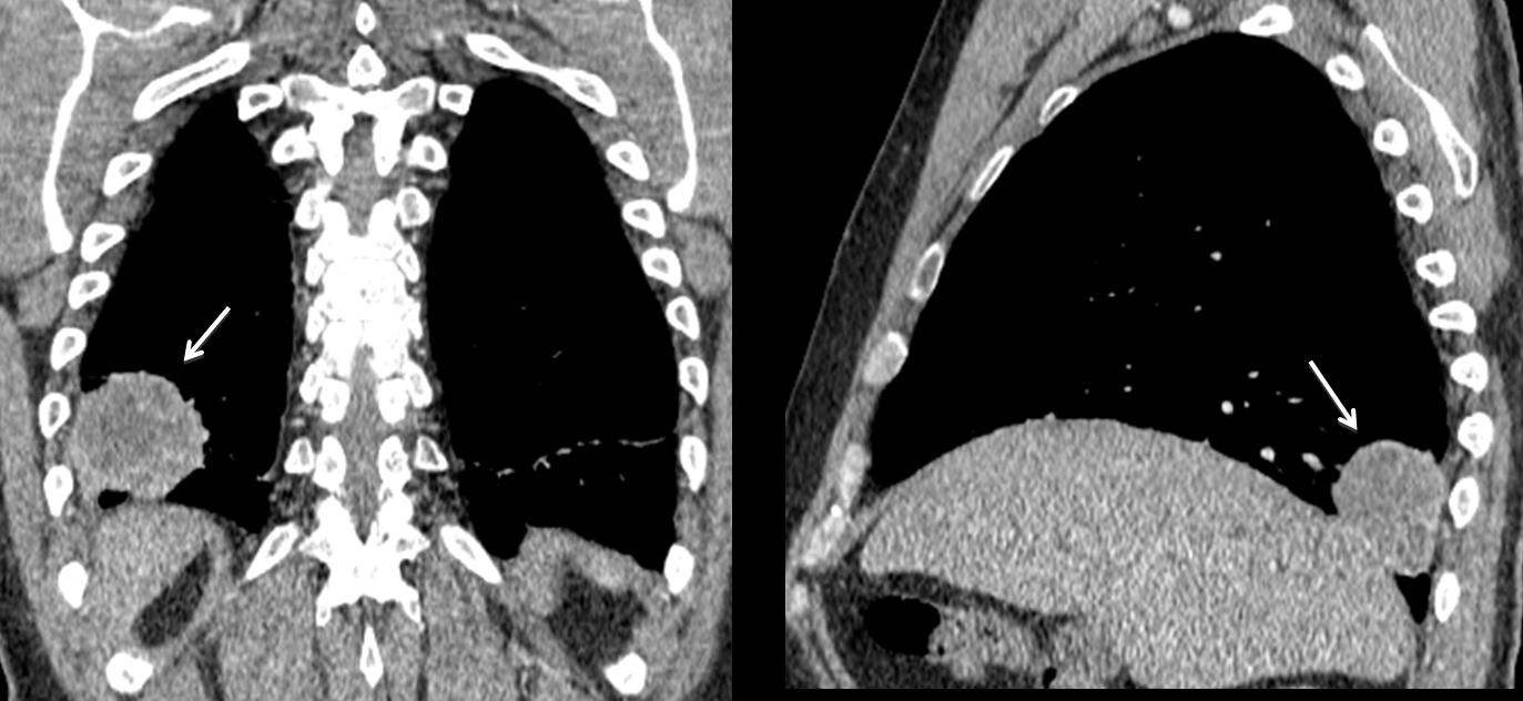
Fig. 4
By far, the most common lesion causing a bulge in the posterior costophrenic sulcus is Bochdalek hernia. Acquired Bochdalek hernia is due to weakening of the diaphragmatic muscle fibres, with protrusion of abdominal fat. The incidence of this condition increases with age and it is not an uncommon finding in asymptomatic elderly individuals.
In Fig. 5, Bochdalek hernia appears as a rounded posterior sulcus lesion (arrows). Note imaging similarity to previous cases.
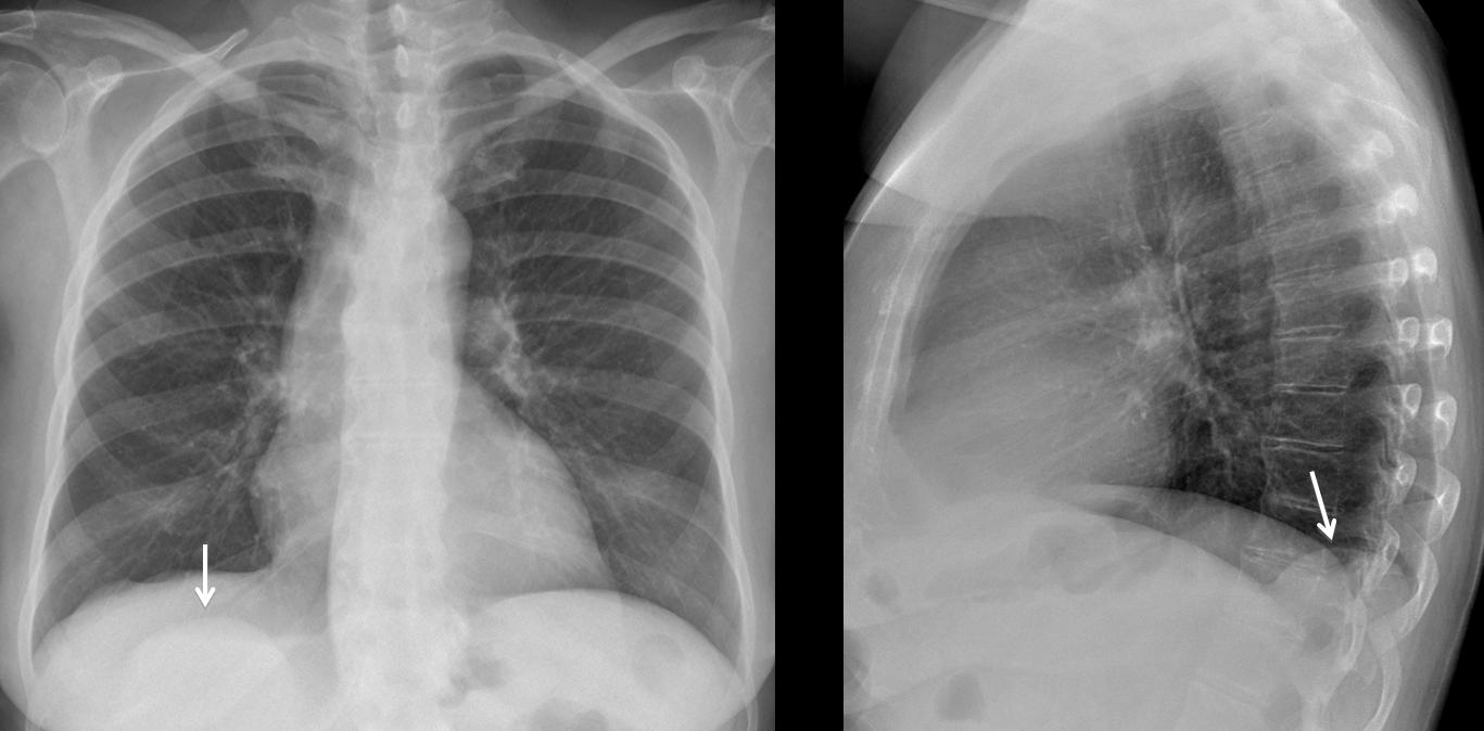
Fig. 5
The diagnosis is confirmed by axial CT, which shows the herniated fat (arrow). Sagittal CT confirms fat herniation through a diaphragmatic hiatus (arrow).
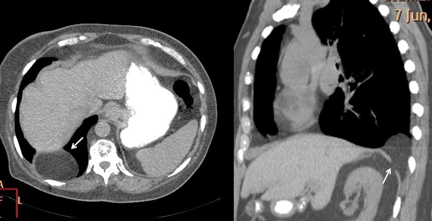
Fig. 6
Bilateral Bochdalek hernia is not uncommon. In this 72-year-old man, chest radiographs show posterior masses in both costophrenic angles (Fig. 7, arrows)
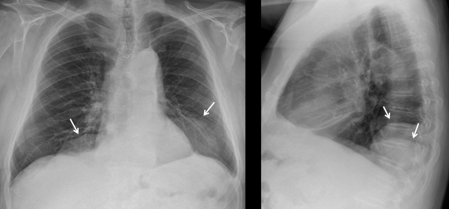
Fig. 7
Coronal and sagittal CT confirm the bilateral herniated abdominal fat (arrows) and posterior discontinuity of the diaphragm (red arrow).
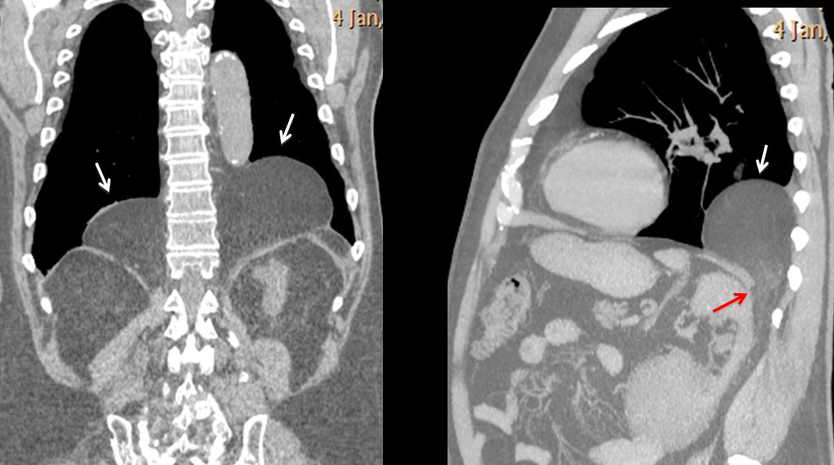
Fig. 8
Diaphragmatic masses are uncommon and cannot be distinguished from other lesions in chest radiographs. A solid mass attached to the diaphragm is likely to be a localised fibrous tumour of pleura. Diaphragmatic cysts are very rare and can be diagnosed by CT. Fig. 9 shows a diaphragmatic bulge (arrows) in a 61-year-old woman that has remained unchanged for the last ten years.
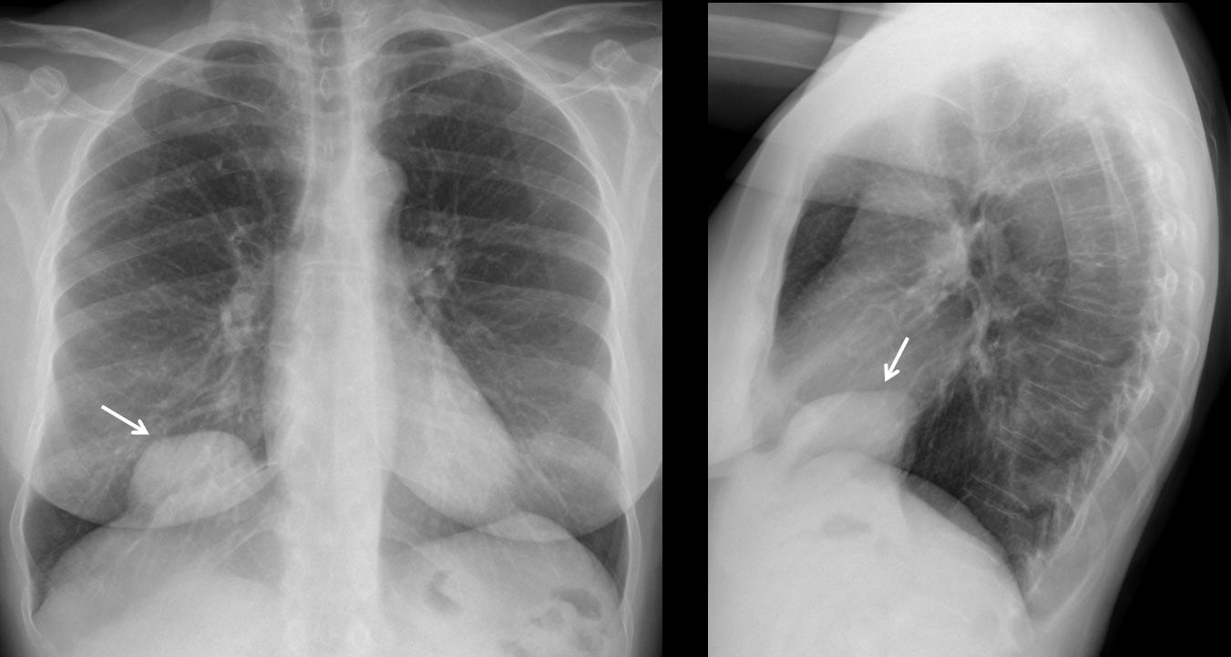
Fig. 9
On unenhanced CT, the mass shows fluid content and peripheral calcifications.
Final diagnosis: diaphragmatic cyst.
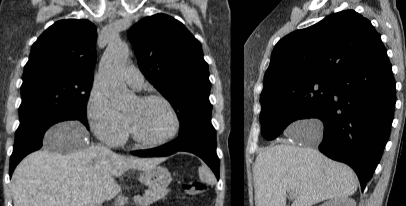
Fig. 10
Follow Dr. Pepe’s advice
Follow Dr. Pepe’s advice:
- Diaphragmatic bulges may be related to a diaphragmatic condition, or reflect disease above or below the diaphragm.
- In case of doubt, CT should be performed to confirm the diagnosis and determine the origin and density of the lesion (fat, fluid, soft tissue).
- Bochdalek hernia is the most common lesion in the posterior costophrenic sulcus, especially in elderly persons.
Recommended reading: Prevalence of incidental Bochladek’s hernia in a large adult population. AJR 177:363-366, 2001
Case prepared by Dr. Pepe


2. Bochladek’s hernia
Bochdalek hernia
The answer is carcinoma of rhe lung
Number 1
Bochdahlik hernia
Round opacity in the right posterior costophrenic sulcus. It could be a posterior mediastinal mass (pleural or neurogenic origin). With the options given I think Bochladek’s hernia is the best. It can be discovered in adults as a small herniation of fat tissue in both sides.
Bochdaleks hernia
diaphragma tear
it could be a diaphragma tear
. Bochladek’s hernia
It looks like bochdalek hernia with gaseous pockets within
I believe the gaseous pockets may be superimposed bowel
There is a strong opinion in favor of hernia and only one for carcinoma. Anybody goes for diaphragmatic cyst?
I do not really like the configuration of aorta. Is he hemodynamiicly stable? I desperately need a chest ct.
No problems with the aorta on CT.
bochdalek’s hernia vs diaphrammatic cyst.. Given the history of trauma diaprhagm tear with subsequent herniation is more likely.
However, Age may favour a diagnosis of carcinoma, but there are no other symptoms or xray findings supportive.
Taking into account that:
1. Muppet is tricky and things could not be what they look at first sight (that is the lesson).
2. Angle of the mass with diaphragma is acute (lateral view) suggesting a supradiaphragmatic origin.
I would choose lung carcinoma.
I may even trickier than you imagine. You may be right and still give the wrong answer. Still, I congratulate you.
Nobody answered diaphragmati cyst, but just analysing the image we cannot say it can`t be. It is rare but diaphragmatic hidatic cyst is an option. In the end, all the options given are possible.
Good. You get first prize!
OK, I give up and wait for the solution.
Muppet is a twisted mind. If the answer is 4, all of the above, Muppet will be barbecued (calçots time).
There is also an osteoporotic wedge shape fracture of a thoracic vertebra. I will go with lung carcinoma with paraneoplasm reaction.
I believe it is carcinoma.patient too old for bochdalek hernia.diaphragmatic cysts r usually between liver and diagram .it is in the chest
Disagree with you. You are thinking of congenital Bochladek hernia. Old people have adquired B. hernia due to the weakening of diphragmatic fibers. Adquired B. hernias increase with age
the lesion is round shape , well defined in the left posterior costophrenic sulcus:
1- despite patient is old age ( goes with tumor), but the lesion is well defined, so this option is unlikely.
2. Bochladek’s hernia, left sided, more in neonates despite some cases reported in adult but there is no findings of herniated bowel in the chest… so it is unlikely.
3. Diaphragmatic cyst, also congenital and more in children , so it is unlikely…
we can noticed that descending aorta is tortuous because of atherosclerotic changes as old age … therefore my differential Dx could be descending aortic aneurysm or hydatid cyst.
Crollo somatico, a cuneo anteriore di T5, fratture costali , con callo osseo , a dx, mentre a sx le fratture costali, sono recenti, senza callo osseo, e riferibili pertanto al trauma in atto.La massa dello sfondato costo-frenico posteriore dx, si raccorda ad angolo acuto con il polmone, indicandone una sede intra-polmonare. Penso allora a lesione maligna polmonare, con metastasi ossee: il trauma toracico ha fatto “scoprire” la patologia di base.
Illustre collega, la tua risposta , ritenuta valida per tutte le opzioni che hai formulato, non mi trovano d’accordo e d’altronde la diagnosi finale è propria quella da me formulata , sulla base della patologia ossea concomitante: vertebrale e costale, dx e sx,con “differente” cronologia nei tempi di comparsa delle fratture costali, con e senza callo osseo. Difronte al caso in esame pertanto la 1 diagnosi è quella da me formulata, salvo a dimostrare la “benignità” delle lesioni ossee concomitanti.Aspetto una tua controreplica.
Patient was operated on and a follow-up chest film a few months later showed healing of the rib fractures. I believe a PET -CT was negative for osseous metastases, although I don´t have the images. Will look for them and send them to you, together with the post-op chest film.
Thank you.
Grazie per la risposta.
The one answer that can be confidently excluded is 4. All of the above. The chance that these 3 pathologies would co-exist is extremely small. I have just looked in PubMed ; it has never been reported.
Perhaps the author meant 4. Any of the above.
You are right. “Any of the above” is the proper wording. My English is getting rusty.
Thanks. I stand corrected