ESR News Quiz Case – February (competition closed)
This month, the prize on offer is free registration for a course offered by the European School of Radiology (ESOR), anywhere in Europe. Simply leave your answer to the question below, as a comment on this post, before February 23 (comments now closed). All correct answers will be entered into a draw and a winner will be selected by the editor of ESR News. The answer and winner will be announced by the end of February.
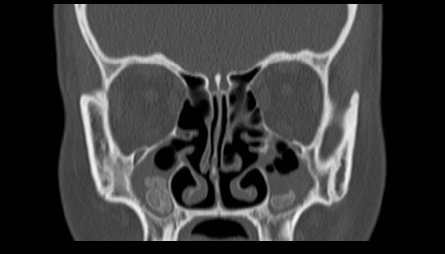
Question: What is the most likely diagnosis?
A. Maxillary antroliths
B. Gardner syndrome
Click here to reveal the answer
Congratulations to the winner of this month’s competition, Helene Gimonet!
Thank you to all participants and well done to those who got the correct answer. Keep an eye out for the March issue of ESR News and your next chance to win registration for an ESOR course of your choice.
Please note that the competition is open to ESR members only. The choice of ESOR course is open, but the winner must fit any eligibility criteria for the chosen course. The prize must be claimed within 12 months.
Click here for a current overview of ESOR courses.


Gardner syndrome
This patient presents hyperdense(calcified/ossified) lesions in both maxillary sinuses that are compatible with osteomas, that may be found in Gardner syndrome. There are also signs of previous operations(i.e. right ethmoidectomy.
Multiple osteomas – B – Gardner’s syndrome
Gardner`s Syndrome (Answer B)
A.Maxillary anteroliths
A.Maxillary anteroliths
A.Maxillary Antroliths
B: Gardners syndrome
Gardner syndrome with paranasal sinus osteomas
Maxillary antroliths
B. Gardner syndrome
B. Gardner syndrome
Gardner’s syndrome
Maxillary Antroliths
A. Maxillary antroliths
B) Gardner’s syndrome
Gardner’s syndrome
Gardner syndrome (B)
A. Maxillary antroliths
A. Maxillary antroliths
Gardner Syndrome
B. Gardner syndrome
B
A. Maxillary antroliths
Answer A
Maxillary antroliths
A. Maxillary antroliths
Maxillary antroliths
A. Maxillary antroliths
A) Maxillary anthroliths
B. Gardner syndrome
A. Maxillary antroliths.
Antroliths can appear solitary or multiple, faintly to extremely radio-opaque masses embedded within the mucoperiosteum. Sometimes with a ‘laminated’ appearance whit radiopaque and radiolucent layers, what can look like layers of an onion. The sinus wall is intact.
Gardner syndrome i guess
B. Gardner syndrome
Maxillary antroliths (correction)
A. Maxillary antroliths
A. Maxillary antroliths
A. Maxillary antroliths
Maxillary antroliths
A. Maxillary antroliths
A. Maxillary anthroliths.