Interview: Dr. Sophie Dellas, head of breast imaging and diagnostics at the University Hospital Basel, Switzerland

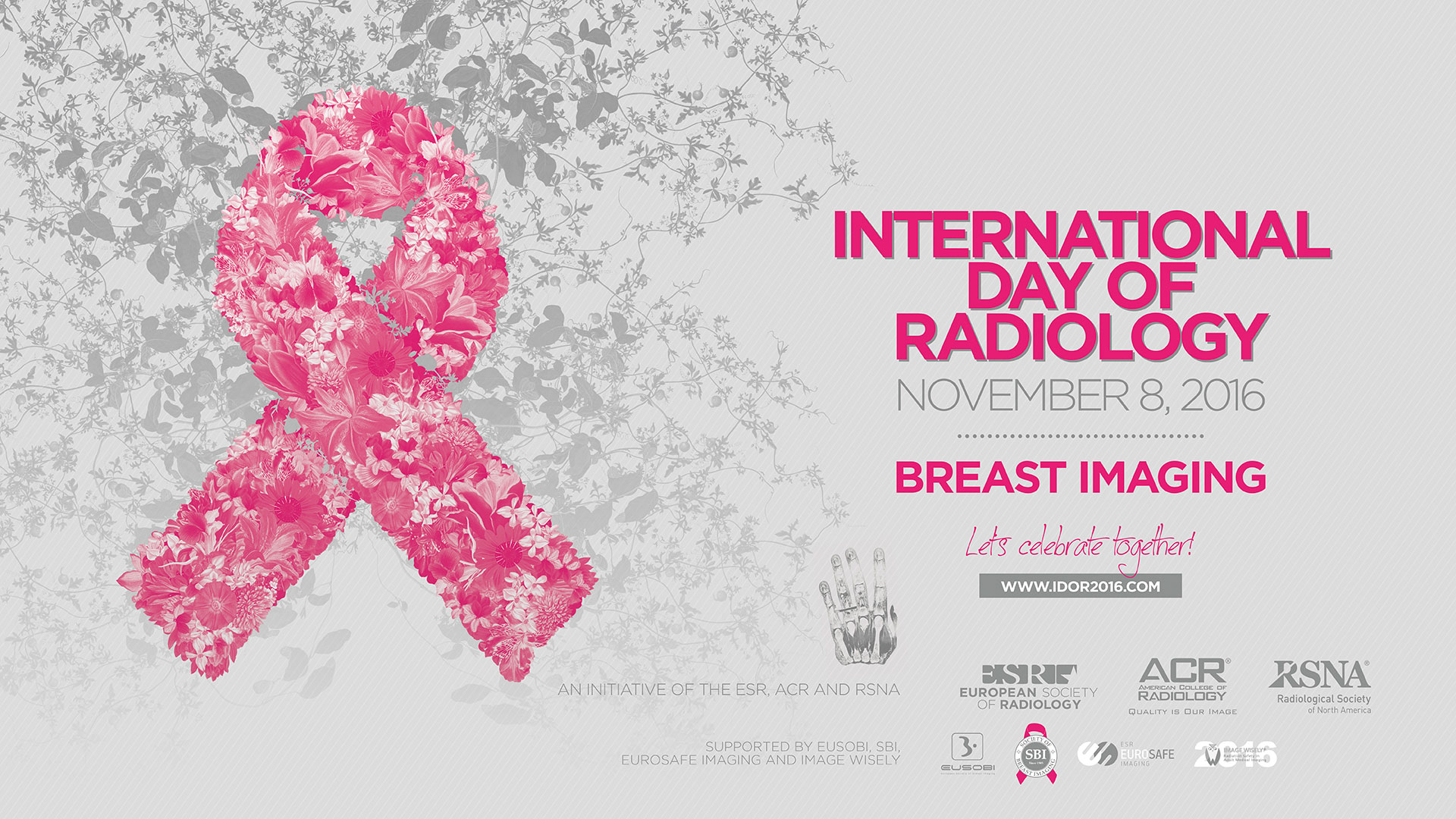
This year, the main theme of the International Day of Radiology is breast imaging. To get some insight into the field, we spoke to Dr. Sophie Dellas, assistant professor of radiology and division head of breast imaging and diagnostics at the University Hospital Basel, Switzerland, and a core team member of the certified breast centre at the same institution.
European Society of Radiology: Breast imaging is widely known for its role in the detection of breast cancer. Could you please briefly outline the advantages and disadvantages of the various modalities used in this regard?
Sophie Dellas: Mammography is the imaging modality of choice for breast cancer screening, but also for diagnosis, evaluation, and follow-up of people who have had breast cancer. Long-term results of randomised controlled trials of mammography screening on average show a decrease in breast cancer mortality of 22% in women aged 50 to 74 years. The main problem of mammography is that it is not a perfect method. Mammography generates 2D images based on the density of tissue for penetrating x-rays. The compression of the breast that is required during a mammogram can be uncomfortable. The compression is necessary to reduce overlapping of the breast tissue. A breast cancer can be hidden in the overlapping tissue and not visible on the mammogram. This is called a false negative mammogram. Mammography is associated with a false negative rate in the order of 10% to 20%. On the other hand, mammography can identify an abnormality that looks like a cancer, but turns out to be normal. This is called a false positive mammogram. Besides worrying about being diagnosed with breast cancer, a false positive means more tests and follow-up examinations. Furthermore, at least some of the cancers found with screening mammography would never otherwise be diagnosed in a patient’s lifetime. The magnitude of such overdiagnosis is a topic of much debate. It is likely to represent up to 10% of breast cancers found on screening mammography and results in potentially unnecessary treatments.

Dr. Sophie Dellas, assistant professor of radiology and division head of breast imaging and diagnostics at the University Hospital Basel, Switzerland.
Breast ultrasound is complementary to both mammography and magnetic resonance imaging (MRI) of the breast. It does not use radiation. It is therefore the initial diagnostic method of choice if breast imaging is required below the age of 40. It allows the confident characterisation of not only benign cysts but also benign and malignant solid masses and the characterisation of palpable abnormalities. The high spatial and contrast resolution of modern breast ultrasound equipment allows the detection of subtle lesions at the size of terminal duct lobular units such as DCIS and small invasive cancers. In women with dense breasts and a negative mammogram, ultrasound therefore is increasingly used as a supplemental screening tool. The major disadvantage of ultrasound as a screening tool is the high risk of false positive findings resulting in unnecessary biopsies. The rate of false positives is much higher with screening ultrasound than with mammography or screening MRI.
Unlike mammography, MRI of the breast does not use radiation. It is safe even though it does require an intravenous injection of a contrast medium. It has a sensitivity exceeding 90% for detecting breast cancer and is superior to mammography and ultrasound. Annual MRI screening is recommended for women with a high lifetime risk of getting breast cancer. Although breast MR imaging is extremely sensitive, its specificity is limited, leading to additional workups and benign biopsies. Good quality breast MR imaging is expensive, time-consuming, and not universally available. Patients with pacemakers, certain aneurysm clips or other metallic hardware, an allergy to contrast agents, or severe claustrophobia are unable to undergo MR imaging.
ESR: Early detection of breast cancer is the most important issue for reducing mortality, which is one reason for large-scale screening programmes. What kind of programmes are in place in your country and where do you see the advantages and possible disadvantages?
SD: Switzerland has no nationwide breast cancer screening programme. There are regional programmes providing organised screening in 12 cantons. The first programmes were established in 1999, the last one in 2014 in Basel. They are organised by the Federation of Swiss Cancer Screening Programmes. Opportunistic mammography screening is the predominant form of breast cancer screening in Switzerland. One major problem is the lack of quality control in the opportunistic screening setting. Opportunistic screening in women at normal risk is not covered by insurance. This is a major problem for women with low income. The health costs of opportunistic screening are high. In 2008 they were calculated to be twice those of organised screening per life year gained.
ESR: Do you know how many women take part (percentage) in these programmes? Do patients have to pay for this?
SD: In 2012 the participation rate of invited women was about 47.8%. There are large differences in participation rates between single programmes. The lowest rate was about 31% and the highest rate about 63% in 2012. The screening mammograms are covered by insurance. Participating women only pay a percentage excess of about 15 to 20 Swiss Francs. Many women get preventive mammograms outside existing programmes. This explains the relatively low participation rate in the existing screening programmes.
ESR: The most common method for breast examination is mammography. When detecting a possible malignancy, which steps are taken next? Are other modalities used for confirmation?
SD: In most cases an additional ultrasound examination of both breasts and axillary lymph nodes will be done. If the initial exams are not conclusive, magnetic resonance imaging may be used as an additional imaging modality to measure the tumour’s size and see how much the disease has grown throughout the breast and to check the other breast for cancer. After completing the imaging tests a breast biopsy has to be done. Usually the breast biopsy is minimally invasive; with a core needle or a vacuum-assisted biopsy, depending on the amount of tissue being removed. Image-guided biopsy is used when a distinct lump cannot be felt, but an abnormality is seen with an imaging test. During this procedure, a needle is guided to the location with the help of an imaging technique, such as mammography, ultrasound, or MRI. A stereotactic biopsy or a tomosynthesis-assisted biopsy is done using mammography to help guide the needle. The result of the biopsy will prove whether an abnormality is cancer. Depending of the type and stage of cancer a treatment plan has to be developed.
ESR: Diagnosing disease might be the best-known use of imaging, but how can imaging be employed in other stages of breast disease management?
SD: The stage of a breast cancer tumour at initial diagnosis determines the prognosis and directs the treatment planning. Current staging guidelines recommend clinical examination and multimodality imaging, which includes conventional mammography, ultrasound of the breast and ultrasound of the axillary fossae for local staging. Systemic radiographic staging evaluation for metastasis is not indicated in asymptomatic patients with normal blood tests and disease-free axillary lymph nodes. Chest x-ray or chest computed tomography, bone scan and ultrasound of the liver to evaluate for distant metastases should be considered in patients with intermediate or high risk of having distant metastasis. PET/CT is increasingly used as a primary staging tool.
Breast MRI plays a role in evaluating tumour response in the setting of neoadjuvant chemotherapy. Information about residual tumour burden assists with preoperative planning and guiding surgical management.
After completion of adjuvant therapy, follow-up focuses on early detection of recurrent disease with the intention of improving long term survival. Mammography plays an important role in surveillance imaging. Most treatment guidelines suggest annual mammography following breast conservation therapy. MRI is useful in women with a history of breast cancer and suspicion of recurrence when clinical, mammographic, or sonographic findings are inconclusive.
PET/CT is appropriate for restaging of breast cancer patients with documented or suspected recurrent breast cancer. CT is used to monitor disease in the metastatic setting.
ESR: How do radiologists’ interpretations help in reaching a diagnosis? What kind of safeguards help to avoid mistakes in image interpretation and ensure consistency?
SD: Breast radiologists have to be familiar with the classification of Breast Imaging Reporting and Data System (BI-RADS) with all available imaging modalities. They have to be trained in reading the images and to use the appropriate imaging modality to guide percutaneous biopsy. They have to read a minimum number of mammograms per year according to the European guidelines, and double reading of mammograms is mandatory. Results of breast biopsies are discussed in multidisciplinary preoperative meetings. The radiologist must assess the adequacy of the tissue sample and evaluate for correlation between imaging findings and histologic results. The decision to recommend surgical excision or appropriate follow-up relies on whether the histologic diagnosis correlates with the imaging findings, a determination that is part of the radiologist’s responsibilities.
ESR: When detecting a malignancy, how is the patient usually informed and by whom?
SD: For the patient, waiting for results can be a real challenge. We try to keep the time between doing the breast biopsy and getting the final result as short as possible. In our breast centre the results of breast biopsies are discussed in preoperative meetings and the treatment plan is developed by the multidisciplinary team. The referring physician gets the radiology report and the pathology report as well as the treatment plan. He tells the patient the results and the next steps to be done.
ESR: How aware are patients of the risks of radiation exposure? How do you address the issue with them?
SD: Modern-day mammography only involves a tiny amount of radiation – even less than a standard chest x-ray. The dose of radiation received during a screening mammogram is about the same amount of radiation a person gets from the natural surroundings (background radiation) in an average three-month period. Breast parenchyma of adolescent girls and young women is most sensitive to a possible carcinogenic effect of radiation exposure. This is one of the reasons why ultrasound is used as the initial diagnostic method if breast imaging is required in women below the age of 40.
ESR: How much interaction do you usually have with your patients? Could this be improved and, if yes, how?
SD: As a breast radiologist doing breast assessment including all kinds of image-guided breast biopsies, I have contact with most of our patients. I tell them what kind of imaging procedure or what kind of biopsy has to be done, why and how this will be done, and about the risks of a certain procedure. Immediately after completion of additional imaging, in cases where there is a suspicious finding, I tell the patient if the result is a normal finding or if further assessment with imaging or an image-guided biopsy is required.
ESR: How do you think breast imaging will evolve over the next decade and how will this change patient care? How involved are radiologists in these developments and what other physicians are involved in the process?
SD: Traditional two-dimensional digital mammography will be supplemented by three-dimensional digital breast tomosynthesis (DBT) and reconstructed 2D mammograms with increased cancer detection rates and reduction in false positives.
But oncologic imaging relies not only on anatomy; it increasingly relies on functional information. Screening and diagnosis of breast cancer needs to use both to improve sensitivity and specificity as well. Contrast-enhanced digital mammography is a test that combines information on vascularity as well as anatomic abnormalities. If further studies show that contrast-enhanced digital mammography is able to depict breast cancers in a fashion similar to that of MR imaging, it could potentially be used in staging breast cancer.
FAST breast MRI techniques may be the standard for breast cancer screening in the future. In a study recently published by Professor Christiane Kuhl and co-authors it was shown that an MRI acquisition time of three minutes and an expert radiologist reading time of less than three seconds are sufficient to establish the absence of breast cancer with a negative predictive value of nearly 100%. Breast MRI provides high resolution images and functional information as well. Unlike mammography, MRI contains information that correlates with proliferation and possibly metastatic potential of a cancer. FAST breast MRI screening, with its capability to detect early neovascularity, could potentially make a big impact on identifying biologically relevant cancers that are currently missed by mammography.
Ultrasound also has the potential to be an ideal screening tool. It is less expensive than MRI and does not require contrast media or ionising radiation. With further development of computing technology automated three-dimensional (3D) whole-breast ultrasound imaging will be available, including multidimensional reformations, reconstructions and tomographic ultrasound. It will replace handheld ultrasound for cancer screening in dense breast parenchyma and in high risk women. Imaging fusion of US information with digital mammography, tomosynthesis or MRI will also be available.
Dr. Sophie Dellas is board certified in radiology. She has spent most of her career at the Clinic of Radiology & Nuclear Medicine at the University Hospital Basel. Her main interest is breast imaging and diagnostics. Dr. Dellas is assistant professor of radiology and division head of breast imaging and diagnostics. She is a core team member of the certified breast centre at the University Hospital Basel and a certified radiologist of the regional screening programme.
Read our interviews with expert breast radiologists from 25 different countries here.


[…] Als Expertin auf diesem Gebiet wurde Frau Dr. Sophie Dellas (Universitätsspital Basel) von der European Society of Radiology zu einem ausführlichen Interview eingeladen. Sie finden dieses höchst interessante Gespräch hier. […]
Hi .Good information.There are some conferences happening in which medical specialty would be Breast Radiology and here is one of those conferences.
Breast Imaging cme – Breast Imaging for Today’s Radiologist is organized by World Class CME and will be held from Mar 30 – 31, 2019 at The Westin Nashville, Nashville, Tennessee, USA. The target audience for this medical event is Radiologists.
For more information please follow the below link:
https://www.emedevents.com/c/medical-conferences-2019/breast-imaging-for-today-s-radiologist-by-world-class-cme
Hi,
Here we provide one Breast Radiology Conference. the conference details are given below.
Around the Clock Imaging is organized by Penn Medicine – Department of Radiology and will be held from Jul 29 – Aug 02, 2019 at Fairmont Pacific Rim, Vancouver, British Columbia, Canada.
For more information please follow the below link:
https://www.emedevents.com/c/medical-conferences-2019/around-the-clock-imaging
Thank you,
Hi,
Thanks for sharing this information.There are some conferences happening in which medical specialty would be Breast Radiology and here is one of those conferences the conference details are given below.
Breast Imaging for Today’s Radiologist is organized by World Class CME and will be held from Sep 28 – 29, 2019 at Gaylord Texan Resort & Convention Center, Dallas, Texas, United States of America. This CME Conference has been approved for a maximum of 15 AMA PRA Category 1 Credit™.
For more information please follow the below link:
https://www.emedevents.com/c/medical-conferences-2019/breast-imaging-for-today-s-radiologist-dallas-1
Hi,
Thanks for sharing this information. There are some conferences happening in which medical specialty would be Radiology and here is one of those conferences the conference details are given below.
Diagnostic and Sub Specialty Radiologists and Radiology trainees are the Target Audience of the conference & it has been approved for a maximum of 20.0 AMA PRA Category 1 Credits™
Source:
https://www.emedevents.com/c/medical-conferences-2019/iu-imaging-update-in-wine-country
Hi,
Thanks for sharing this information. There are some conferences happening in which medical specialty would be “Breast Radiology” and here is one of those conferences the conference details are given below.
World Class CME: Breast Imaging for Today’s Radiologist 2020 is organized by World Class CME and will be held from Sep 19 – 20, 2020 at Las Vegas, Nevada, USA. This CME course has been accredited for a maximum of 15.0 AMA PRA Category 1 Credit™.
Source: https://www.emedevents.com/c/medical-conferences-2020/breast-imaging-for-today-s-radiologist-las-vegas
Hi,
Thanks for sharing this information. There are some conferences happening in which medical specialty would be “Primary Care” and here is one of those conferences the conference details are given below.
Breast Radiology: 2021 Breast Ultrasound With Tom Stavros CME is organized by World Class CME (WCC) which
will be held from Jan 15 – 17, 2021 at San Antonio, Texas, USA. World Class CME designates this live activity for a maximum of 21 AMA PRA Category 1 Credits.
Source: https://www.emedevents.com/c/medical-conferences-2021/breast-ultrasound-with-tom-stavros-2021