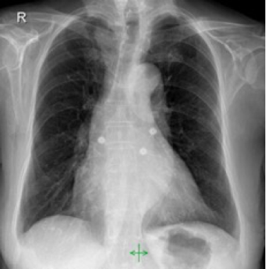
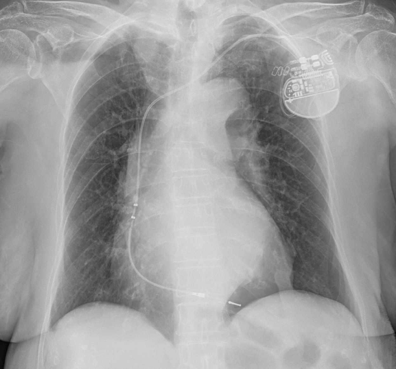
This is a 55 year old patient and the AP radiograph was performed as a control after a pacemaker placement.
Final solution:
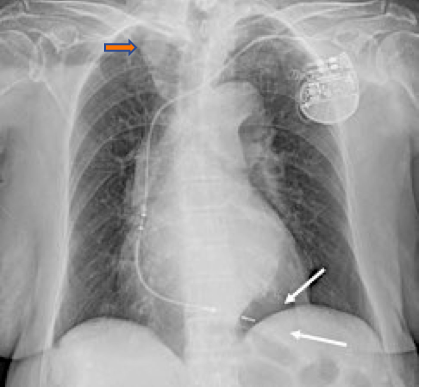
Regarding this case some of you thought that there was a right superior mediastinal “mass”, but the convex image you see, disappears at the level of the clavicle (orange arrow), therefore is anterior ( Cervico-thoracic sign) and in a person with clear vascular elongation ( see the descending aorta ) this represents the supra-aortic vessels mainly the subclavian artery.
I must congratulate “Fingers crossed” because indeed he or she saw the lucent area under the heart (arrows).
In the Chest X Ray obtained before the pacemaker was implanted this black area is nor present.
This sign is typical of Pneumothorax, especially in supine radiographs where it tends to accumulate in the lower thorax.
24 hours before
After pacemaker placement
Pneumothorax is a common complication of pacemaker placement through the subclavian vein. Up to 10 % may develop a pneumothorax but only 1% will require drainage.
The supine radiograph poses a challenge for the detection of pneumothorax since the air will collect near the abdominal region and the heart.
Several signs of pneumothorax in the supine position have been described.
Some of the signs cited by Radiopaedia are::
Lucency in one hemithorax
Deep sulcus sign
Increase sharpness of heart diaphragm and mediastinal structures
Double diaphragm sign
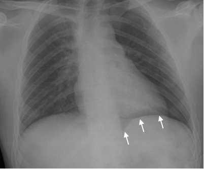
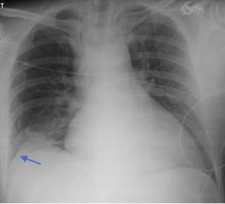
In this other ICU patient air is collected under the heart and above the diaphragm. (White arrows). An upright radiograph confirmed the pneumothorax. (Black arrows).
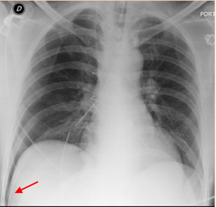
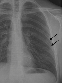
Portable chest film showing a deep sulcus sign (red arrow) after several attempts to place a subclavian line. Two days later, after chest tube placement, the pneumothorax has resolved and the right costophrenic sinus has returned to normal (blue arrow).( from J. Caceres)
Points to remember:
1 – Pneumothorax is a common complication of pacemaker placement.
2 – In supine chest radiographs look for dark areas in the lower thorax, near the heart, diaphragm and costophrenic sulcus.


Pneumothorax in the left lower region?
Shorter electrode in SCV instead of right atrium?
Besides the increased vascular markings, enlarged heart etc.
Upper mediastinal mass
Upper mediastinal mass (right side)
There s a paravertebral opacity on the left side, it cold be aortic pathology
Mass in right upper zone
why the photo can occur?
RV perforation plus pericardial effusion?
I feel good!
And I’m a he!