José Vilar and Friends Case 40 (Update: Solution + Points to remember!)
Dear friends,
another case to test your ability to detect lesions.
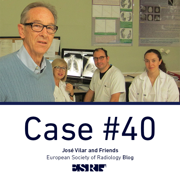
The patient is a 68 year old woman. The finding was just incidental.
Where is the pathology and what could it be? Check the images below.
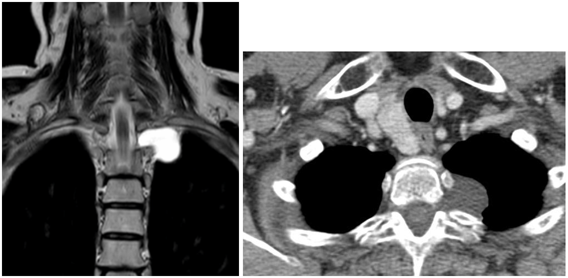
Hello, it seems that the case is either too easy or too difficult. Here are a couple of additional images that can solve the problem.
Diagnosis?
Click here for the answer
This case was visited by 2.976 viewers but, for some mysterious reason, only one brave person made a very close diagnosis ( Suvinay Saxena congrats!)
Let us see, dear friends, what are the findings and what we can extract from the case.
The Chest radiographic study shows an abnormal convexity in the left apex (arrow). The lesion is well defined and convex towards the lung, therefore is extra pleural. Since the lesion is above the clavicles it has to be posterior; the lungs seen in the PA above the clavicles are posterior.
Now look at the lateral projection and you will see the lesion also, well-defined, and superposed to the spine. (Arrow)
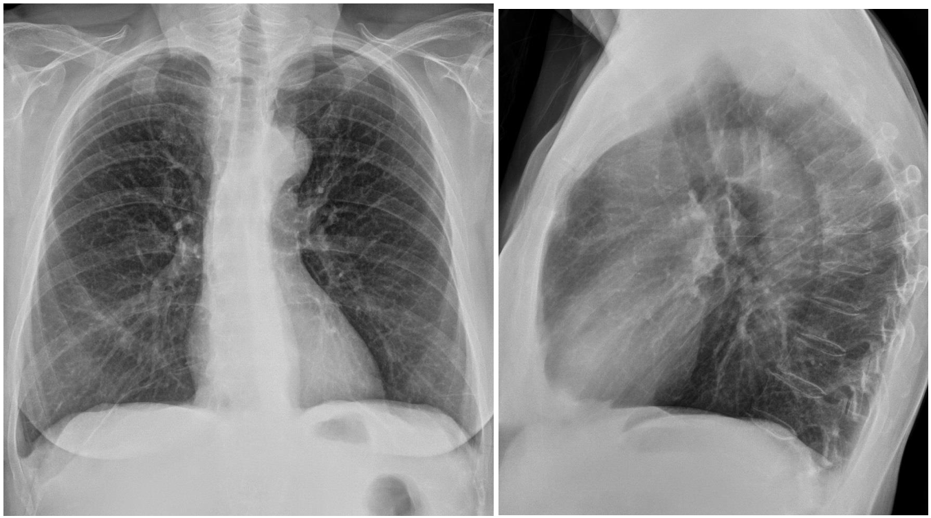
We are then dealing with a posterior mediastinal lesion. The next step will be to obtain a CT with contrast.
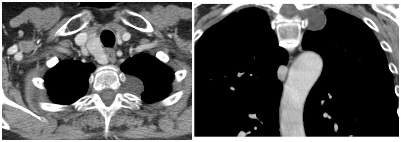
The images, axial and coronal, show that the lesion is related to the spinal canal and has a water density, thus is cystic. The lesion is well defined, water density, has no solid component and there is no contrast uptake. These features characterize a mediastinal lesion as pure cystic.
Additional MR images show similar findings regarding the lesion, but in the myelography sequence other smaller cysts are seen.
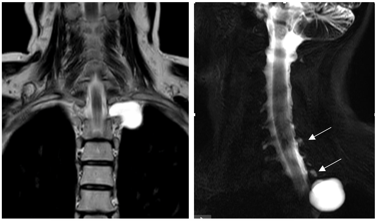
Since most posterior mediastinal lesions have a neural origin the diagnosis is not too difficult in this case, especially since a clear origin in the canal is seen. The diagnosis is then: spinal meningeal cyst.
Points to consider:
- Chest radiography depicts quite often mediastinal lesions as long as the lesion is in contact with the lung.
- Remember that lesions see above the clavicles in the PA projection are posterior.
- CT with contrast is the technique to follow when a mediastinal lesion is detected in chest radiographs.
- If the lesion is cystic always make sure there is no solid component or contrast uptake. Tumours may be partly cystic.
- Meningoceles may be related to neurofibromatosis or just incidental and are not treated if asymptomatic.


Neurogenic tumor- schwanomma. DD neurofibroma
pa view of radiography shows well defined radioopacity in left upper zone with convexity towards lung. so likely in mediastinum with lateral radiograph confirming posterior mediastinal lesion. differetial goes in favour of neurogenic lesion. confirmed on ct and mri as spinal meningeal cyst