José Vilar and Friends Case 41 (Update: Solution + Points to remember!)
Friends, the last case was seen by many, but only one person dared to try a diagnosis.
Let me see this time if you become more implicated. This is a case from my files. (Hospital Universitario Doctor. Peset. Valencia).
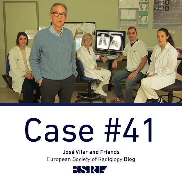
This is a 65-year-old lady with long standing cardiac pathology.

The patient has clear cardiomegaly and other signs of cardiac insufficiency,
But… do you see anything else?
Update: Additional images
The image shows, as Anastasia indicates in her post, a round lesion projected over the left heart and located posteriorly. The lesion does not seem to be in the lung. Then, if it is not in the lung why is it quite well defined especially in the PA projection?
The answer soon.
Click here for the answer
Well, in the last minute Swarnadhanu made the correct diagnosis. But let me explain what are the findings.
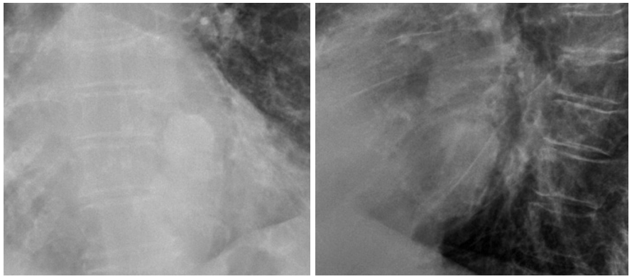
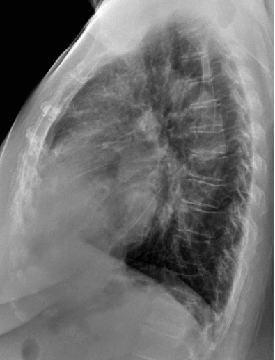
The chest radiographs show a rounded density in the posterior aspect of the heart. The lesion is not in the lung, but is well seen because its density is higher than that of the soft tissues. Therefore, the reason why we can see the lesion is because it is calcified.
The location of the lesion indicates a cardiac origin. Calcifications in the heart are common in elderly people. Linear calcifications are usually vascular (Coronaries, aorta and branches) but may be also due to calcium in the wall of the cardiac chambers or in the pericardium. In our case this is a rounded calcification and according to the location it fits perfectly with the mitral valve.
Two types of calcifications of the mitral valve annulus can be seen frequently:
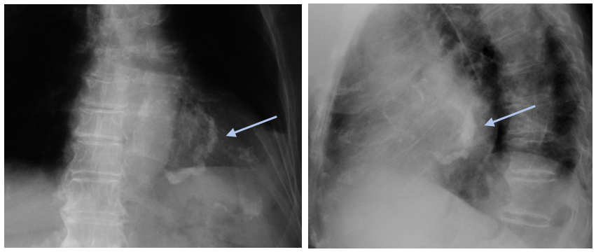
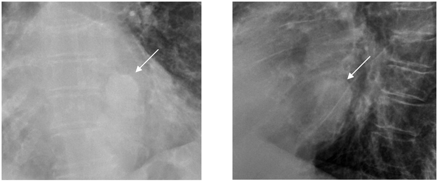
- degenerativemitral annulus calcification and as is seen in this example (blue arrows)
- Liquefactive necrosis or caseous mitral valve calcification as in our case. This entity is a variant of the more common mitral annulus calcification, in which there is a fluid like necrotic component (tooth paste like). It is a benign condition although it may accompany mitral stenosis, and or insufficiency.
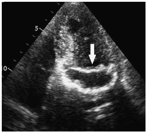
It has been described especially by ultrasonographers as a echogenic rim with a central lucency that can be mistaken for an abscess or tumour.
(Anxo Martinez. Radiología:2010)
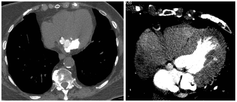
In CT the calcification of the mitral valve annulus appears thick and bulky . Observe that in the contrast image the calcification is partially hidden and more difficult to interpret.
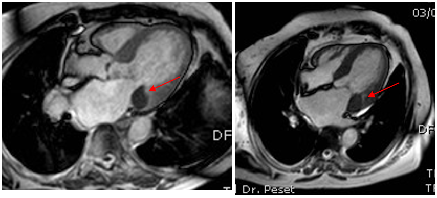
In MRI liquefactive necrosis will appear with very low signal intensity. (Red arrow)
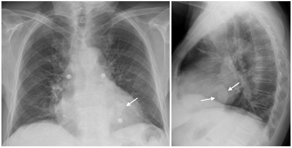
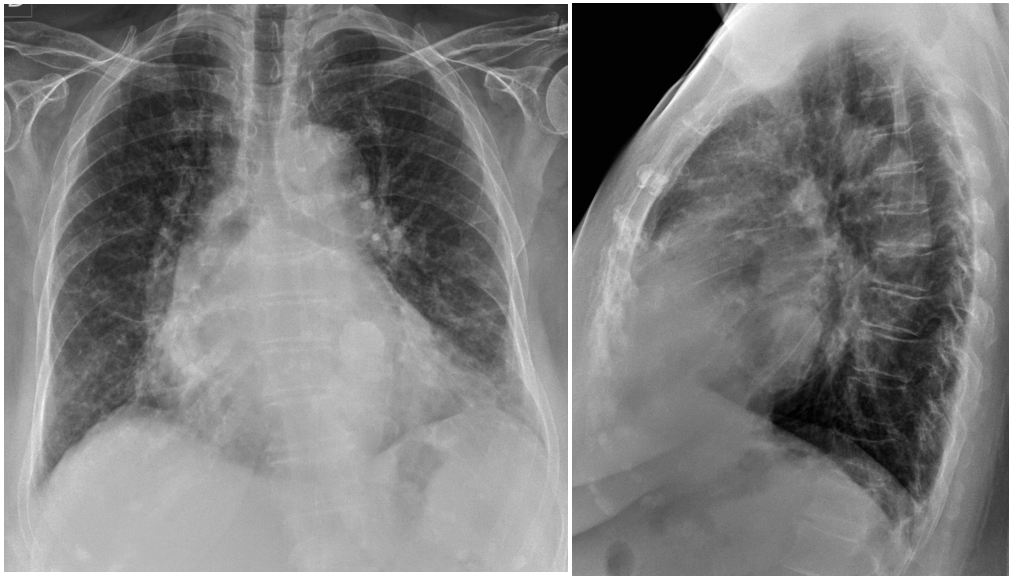
In chest radiographs this entity can be suspected when we see a rounded dense lesion in the area of the mitral valve. In the lateral projection it has often an oblong shape as in this other case. (Arrows).
Main points:
- Calcification of the mitral valve fibrotic annulus is common and usually asymptomatic.
- Caseous or liquefactive necrosis of the annulus is rare and has usually no clinical relevance, but may be mistaken for other conditions.
- The chest radiographic study with two projections may suffice to make the diagnosis of mitral caseous necrosis, and help when ultrasound, CT or MRI show cardiac confusing findings.
Post Data: Some radiographic findings help to avoid confusion and may eliminate unnecessary examinations. All it takes is to read your radiographs and know the basics.


There is a soft tissue mass located at the left retrocardiac space – lower mediastinum, that appears to have wall. Differential diagnosis of epiphrenic diverticulum and esophageal duplication cyst.
Well seen Anastasia but keep thinking…
possibilities of the retrocardiac space mass calcification
mitral valve and annulus calcification.
left ventricular wall calcification
left atrial myxoma calcification
Differential could include left ventricular aneurysm.