Dear Friends,
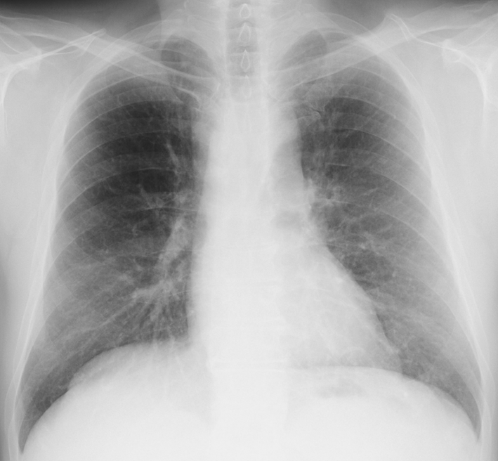
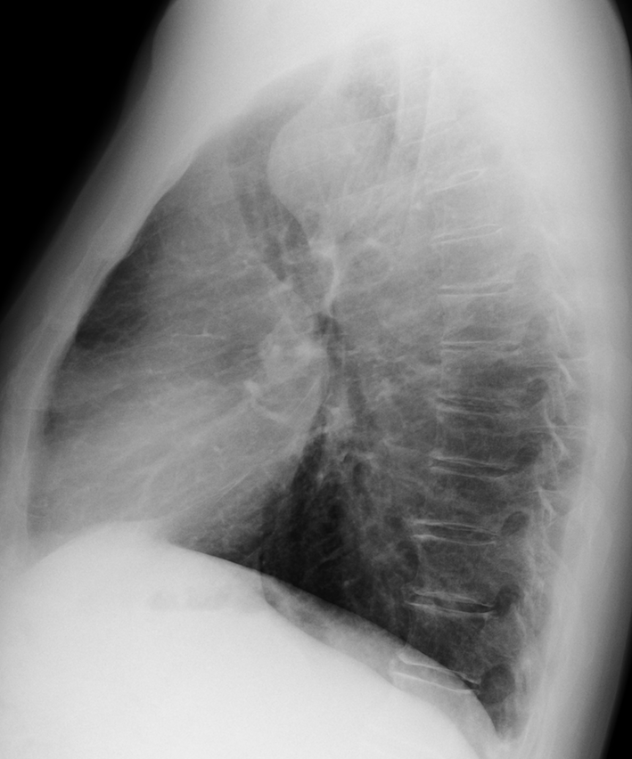
Today’s images belong to a 63-year-old smoker with a persistent cough.
What do you see?
Check the images below, leave your thoughts in the comments section, and come back on Friday for the answer.
Click here for the answer
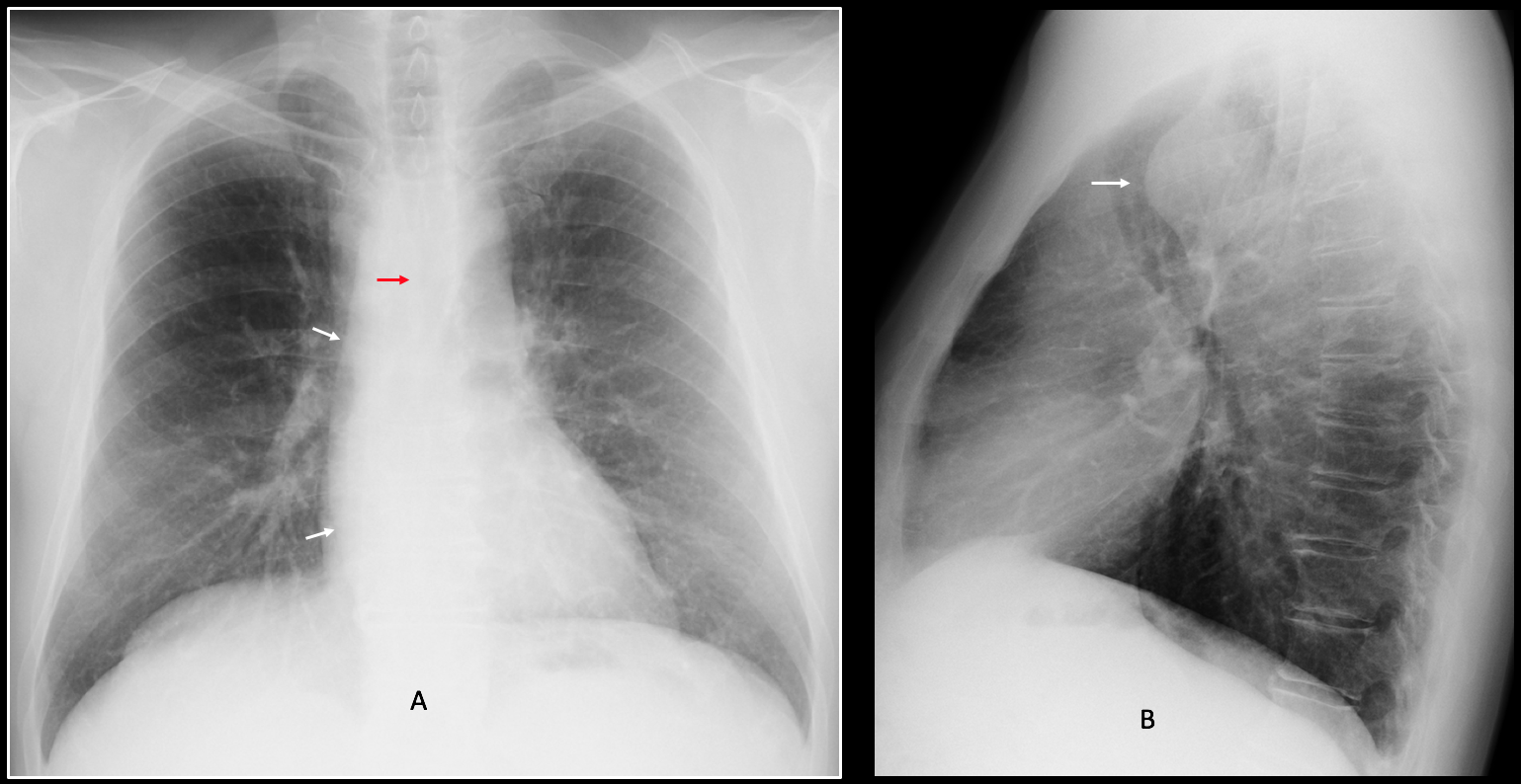
Findings: the most obvious finding is a retro-tracheal mass in the lateral view (B, arrow). The answer to the etiology of the mass lies in the PA view, which shows an imprint in the right wall of the trachea (A, red arrow) and the descending aorta on the right side (A, arrows). In a patient without significant symptoms the most likely diagnosis is a right aortic arch.
Coronal and axial enhanced CT confirms the diagnosis of right aortic arch with a Kommerel diverticulum (C-D, arrows) that explains the anterior tracheal displacement.

Final diagnosis: right aortic arch with Kommerel diverticulum
Congratulations to Diogo, who was the first to suggest the correct diagnosis
Teaching point: remember that right aortic arch occurs in up to 0.1% of the population. A posterior tracheal imprint in an asymptomatic patient should prompt a search for a right aortic arch in the PA view.


There is a subtle widening of the mediastinum on the PA view, and a rounded opacity on the lateral view displacing the trachea anteriorly ( middle mediastinal lesion).
The is also a small retrosternal consolidation.
Impression: middle mediastinal mass (developmental cyst, lymphadenopathy, vascular lesion, neurogenic tumor..).
The retrosternal lesion might be a tumor or infection.
There is a large mass displacing the trachea anteriorly. A dilated anomalous right subclavian artery (Kommerell diverticulum) would be a nice guess.
Hello!! There is a medium mediastinal lesion that displaces anteriorly the trachea. Perphaps a vascular anomaly: double aortic arc vs aberrant subclavia with kommerell´s diverticulum?
Hello Professor,
clearly we are dealing with a mediastinal abnormality and as muppet always says mediastinum is mostly composed of vessels 🙂
– on lateral view there is quite big opacity in middle mediastinum, shifting the trachea to the front
– below that, just above the carina there is (or at least I think there is ;P) also more subtle modeling of the trachea from the front
– on front cxr i don’t see a typical aortic knob on the left side, suggesting RAA, but there is some shadow crossing the tracheal line just above carina – my bet is on double sided aortic arch
Hello, big fan! Nice discussion. Glad you pay attention to Muppet teachings. He is elated!