ESR Rising Stars Speakers at ECR 2012
Welcome to the seventh and last installment of our series introducing our Rising Stars, who submitted the best student abstracts and have been invited to present their work at ECR 2012 in front of a real congress audience. Each speaker will be announced individually on the Rising Stars Facebook page, with a more detailed look, including their full abstracts, here on the ESR Blog. Our speakers Ilaria Merli, and Maria Bercea will close the presentation of this year’s speakers:
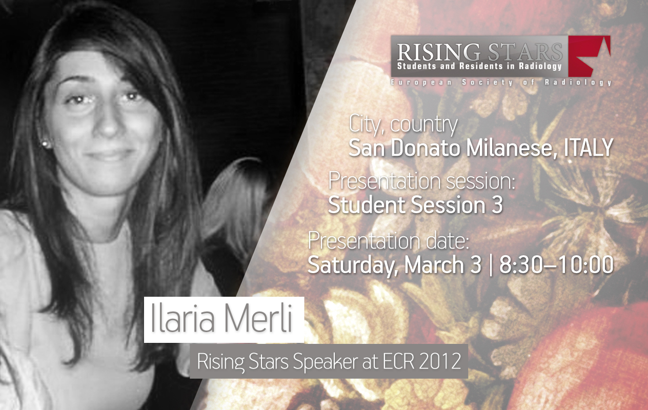
ECR 2012:
Student Session 3 | March 3, 8:30 – 10:00
CV (excerpt):
Education/Profession:
Studying Medicine at the University of Milan, Italy
Abstract:
Which one of these was my first x-ray? The first one I ever saw when I was in the ward of pulmonology or the one I saw during my first radiology lesson? Neither: it’s the first one I actually felt was mine. My first x-ray was a brain CT scan; I was asked to report it as an exercise. I usually follow the radiologist as he/she reports images and every day I try to improve my skills in detecting findings. This opportunity to report like a real radiologist gave me the ability to check what I had learnt during the period I spent in the Unit of Neuroradiology. I understood that the saying ‘you can only see what you know’ is true. If you are sitting near the radiologist, everything seems to be easy to find. When you are alone with the image you can only count on your own knowledge. When I started looking at the images I was immediately impressed by an area of hyperdensity in the right temporal lobe. I was so excited by my discovery of the lesion that I was dazzled by it and I didn’t see a small hyperdense area in the left cerebellar hemisphere. The correction of my report’s proof showed me my mistake. Because of this experience, I now understand that it’s important to focus on every centimetre of the image, without being influenced by previous findings. This approach to the radiological image is crucial in order to avoid making a blunder.
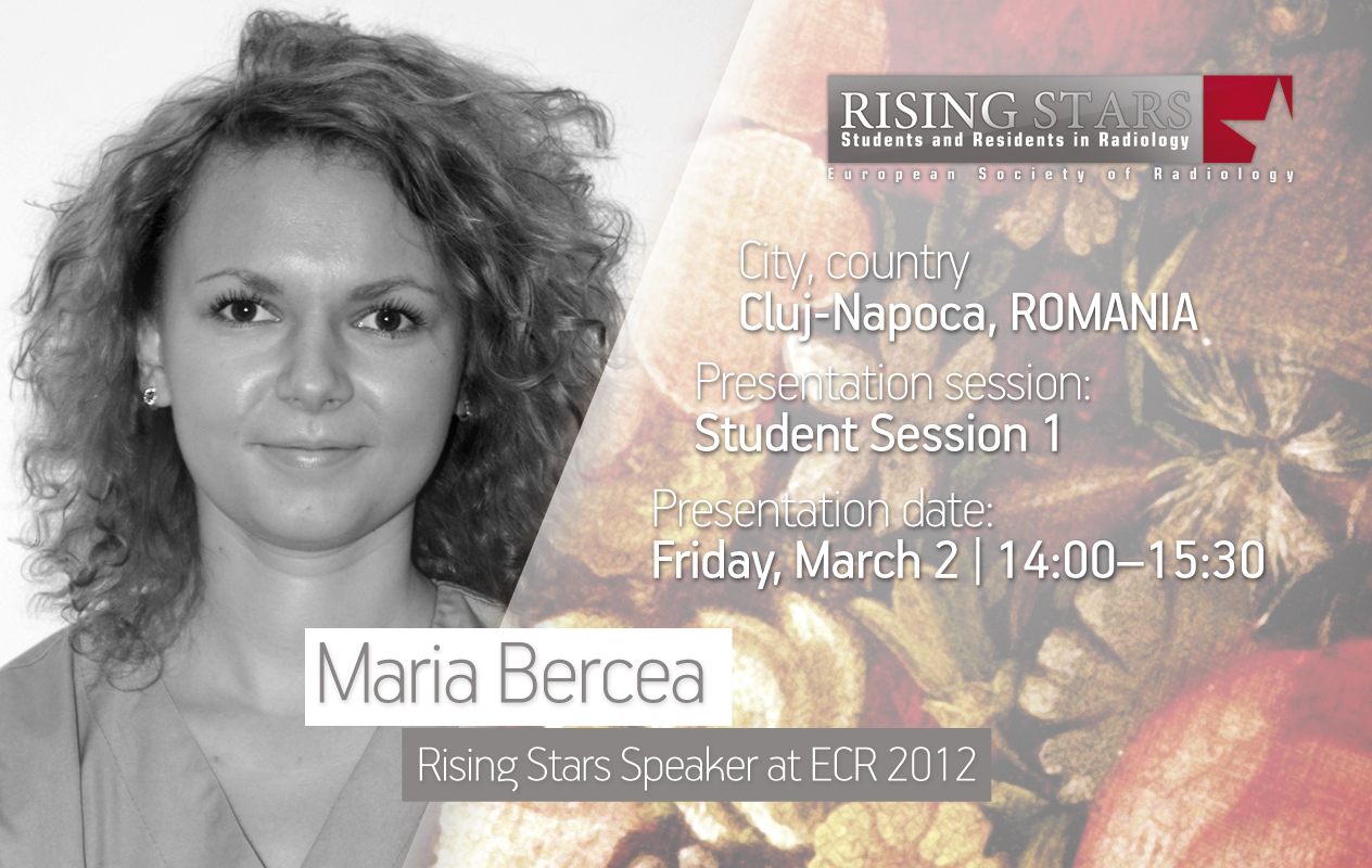
ECR 2012:
Student Session 1 | March 2, 14:00 – 15:30
CV (excerpt):
Since 2006 Studying at the Faculty of Medicine, Iuliu Hatieganu University of Medicine and Pharmacy, Cluj-Napoca,
Abstract:
RADIOLOGIST 2051: What will a radiologist’s day-to-day work look like 40 years from now? After reading several essays by many noted radiology specialists of our time, on how they would like radiology to improve, I have assembled some of their ideas and I have come up with a view of the future of radiology. The Radiologist of 2051 will be a sub-specialist and will manage the new information flow while keeping up with new discoveries in his sub-specialisation. He/she will supervise over the patient through a protective screen while calculating a personalised radiation dose for the procedure and patient. Once the image is obtained, the protective screen will turn into a widescreen touch panel where the radiologist can see the image in full size or even larger and can observe the abnormalities which he can then highlight. He/she will be able to upload the image to an enormous global image data base that analyses and compares his/hers with others that have the same pathology. Through a social network he/she will be able to get in touch with the current doctor who can answer all the questions he/she has about the patient’s clinical past. Afterwards, proceeding towards the final diagnosis the doctor will be able to access all the radiologists on call at the time, who are online, and ask their opinion. These are a just few things which, if implemented, could make the future of radiology even brighter than it already is.

