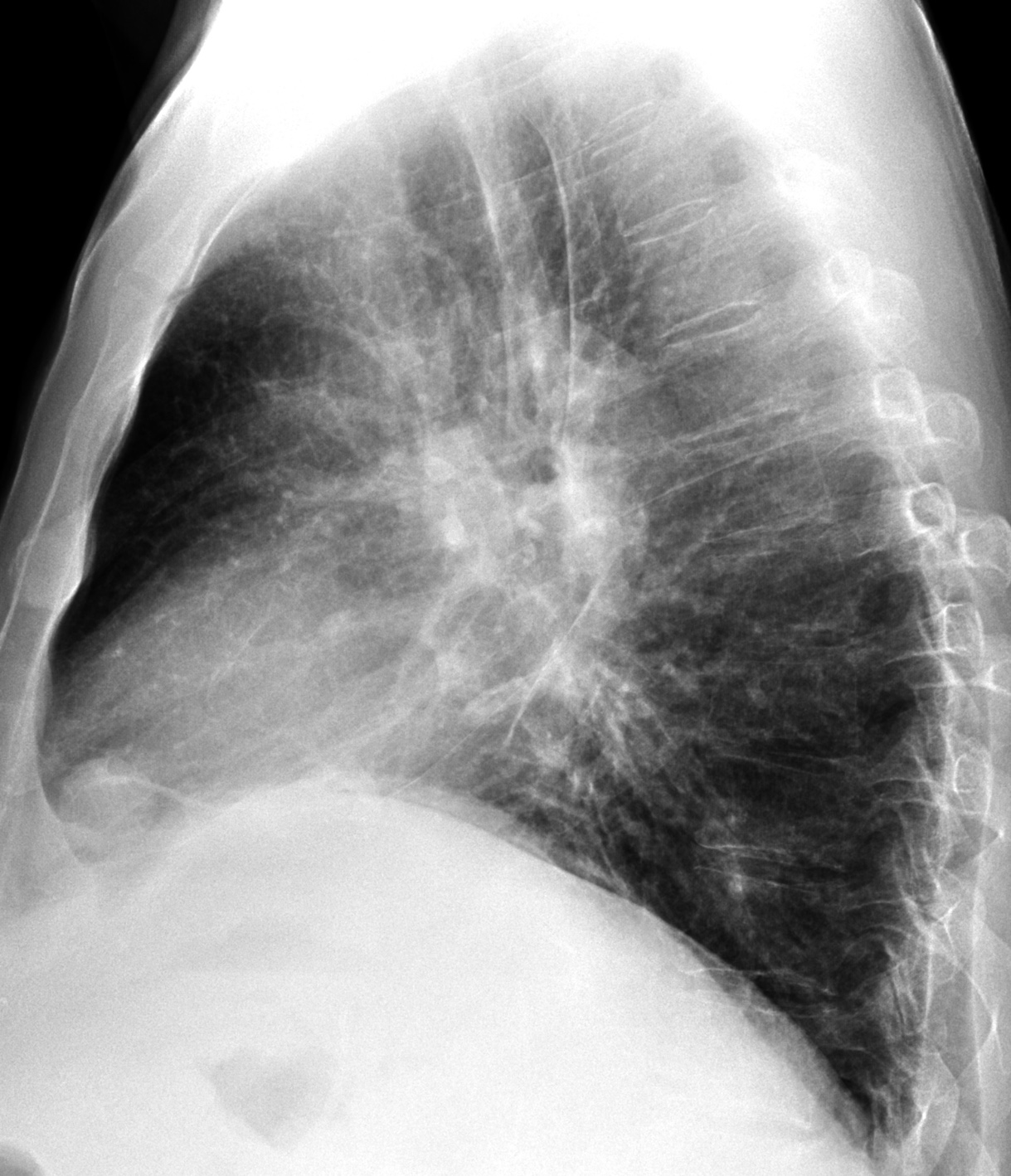
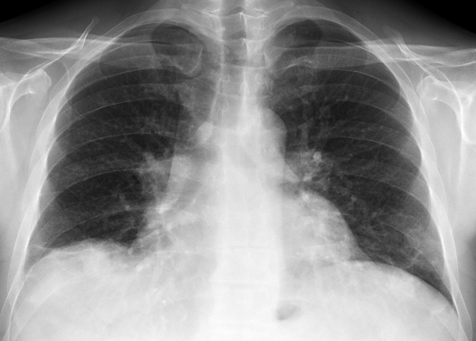
Today I am showing chest radiographs of a 52-year-old man with liver cirrhosis and moderate dyspnoea. What do you see?
Check out the images below, leave your thoughts in the comments section, and come back on Friday for the answer.
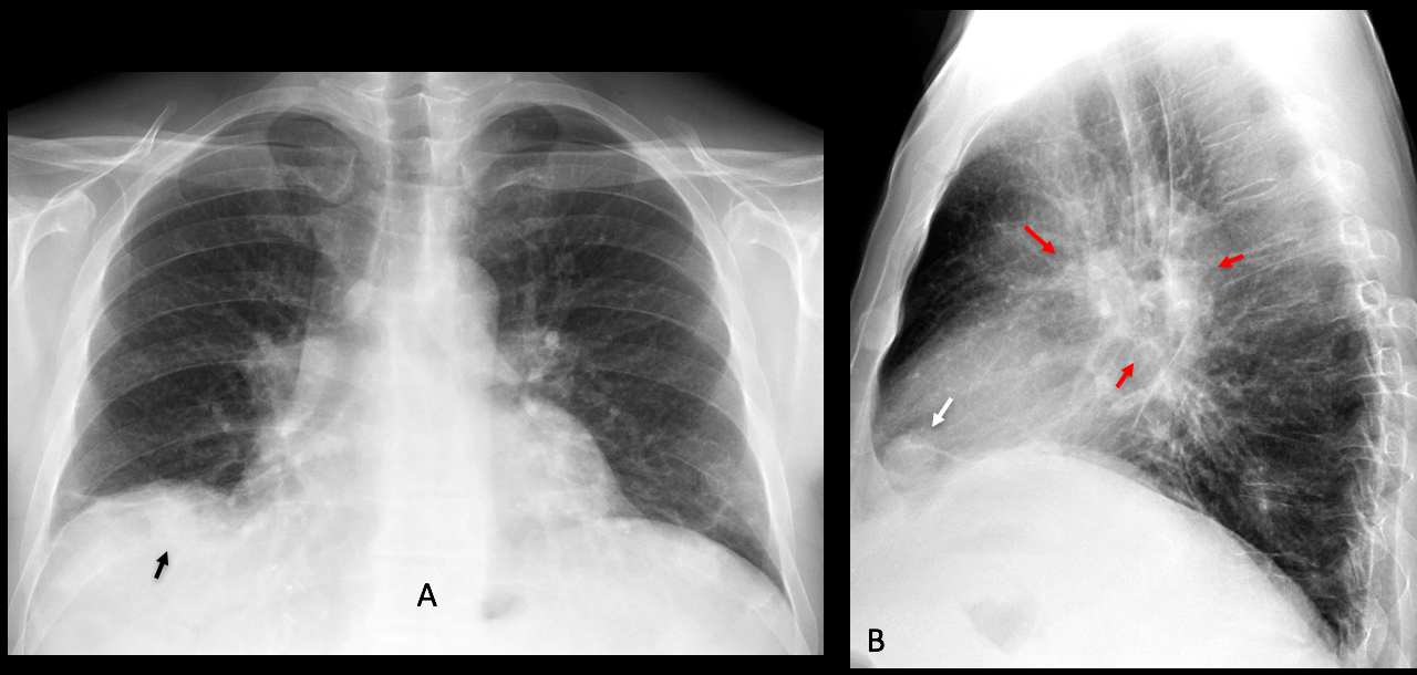
Findings: the main findings are visible in the lateral radiograph, which shows a well-defined pulmonary nodule in the anterior costophrenic sinus (B, arrow). The hila are prominent, with a donut sign (B, red arrows). The nodule is hidden behind the right hemidiaphragm in the PA view (A, arrow).
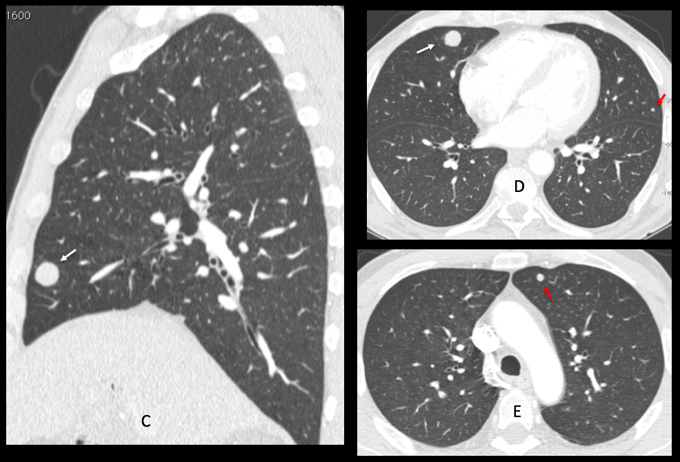
Sagittal and coronal CT confirms the nodule (C-D, arrows) as well as other nodules (D-E, red arrows).
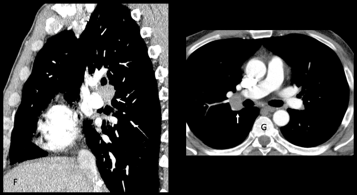
Enhanced sagittal and axial CT show enlarged lymph nodes in the right hilum (F-G, arrows). Needle biopsy of the main nodule confirmed metastases from hepatocarcinoma.
Final diagnosis: pulmonary and hilar metastases from hepatic carcinoma
Congratulations to Alexander Quirós, who was the first to suggest the diagnosis.
Teaching point: value of the lateral film (nodule and hilar lymph nodes).