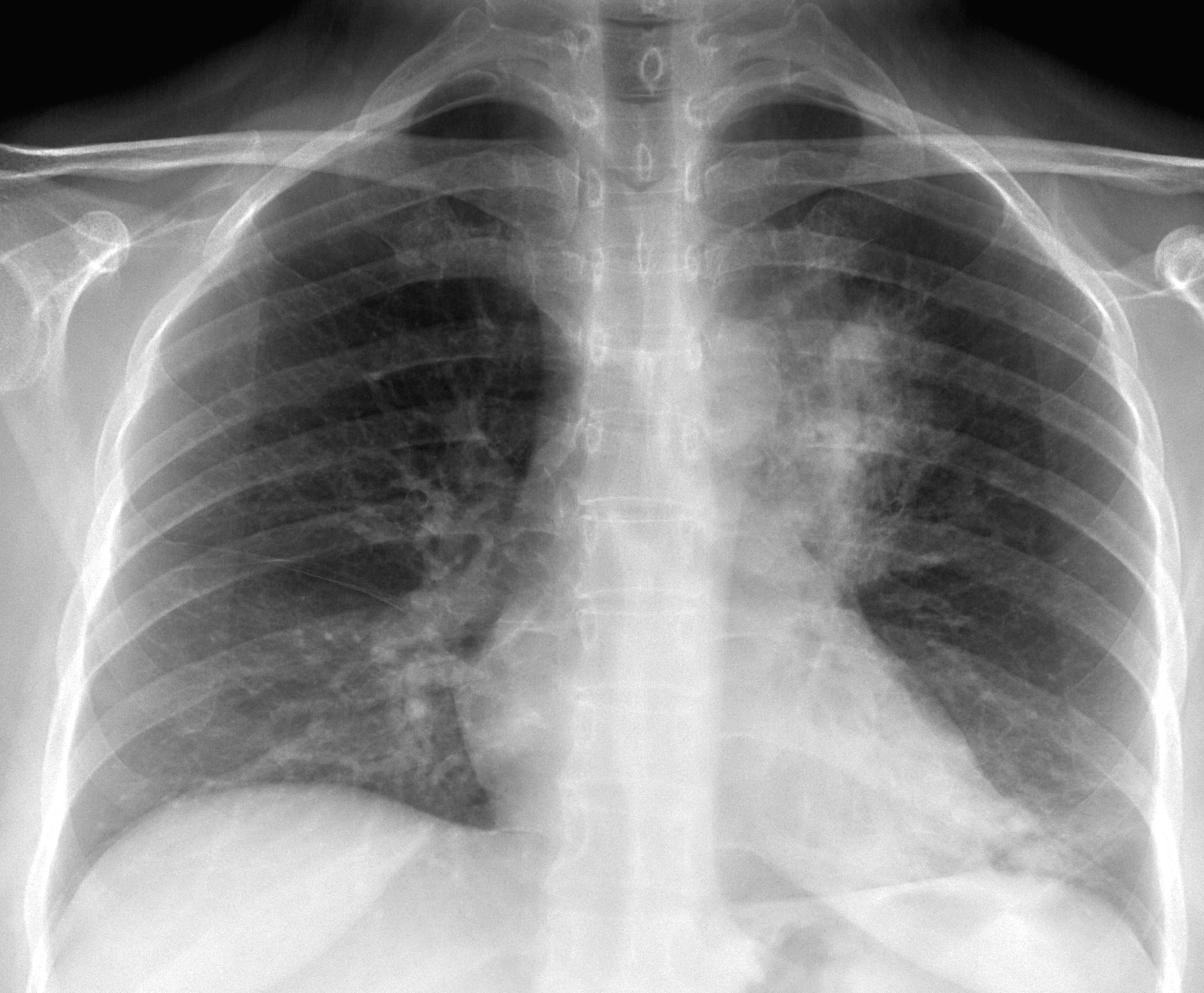
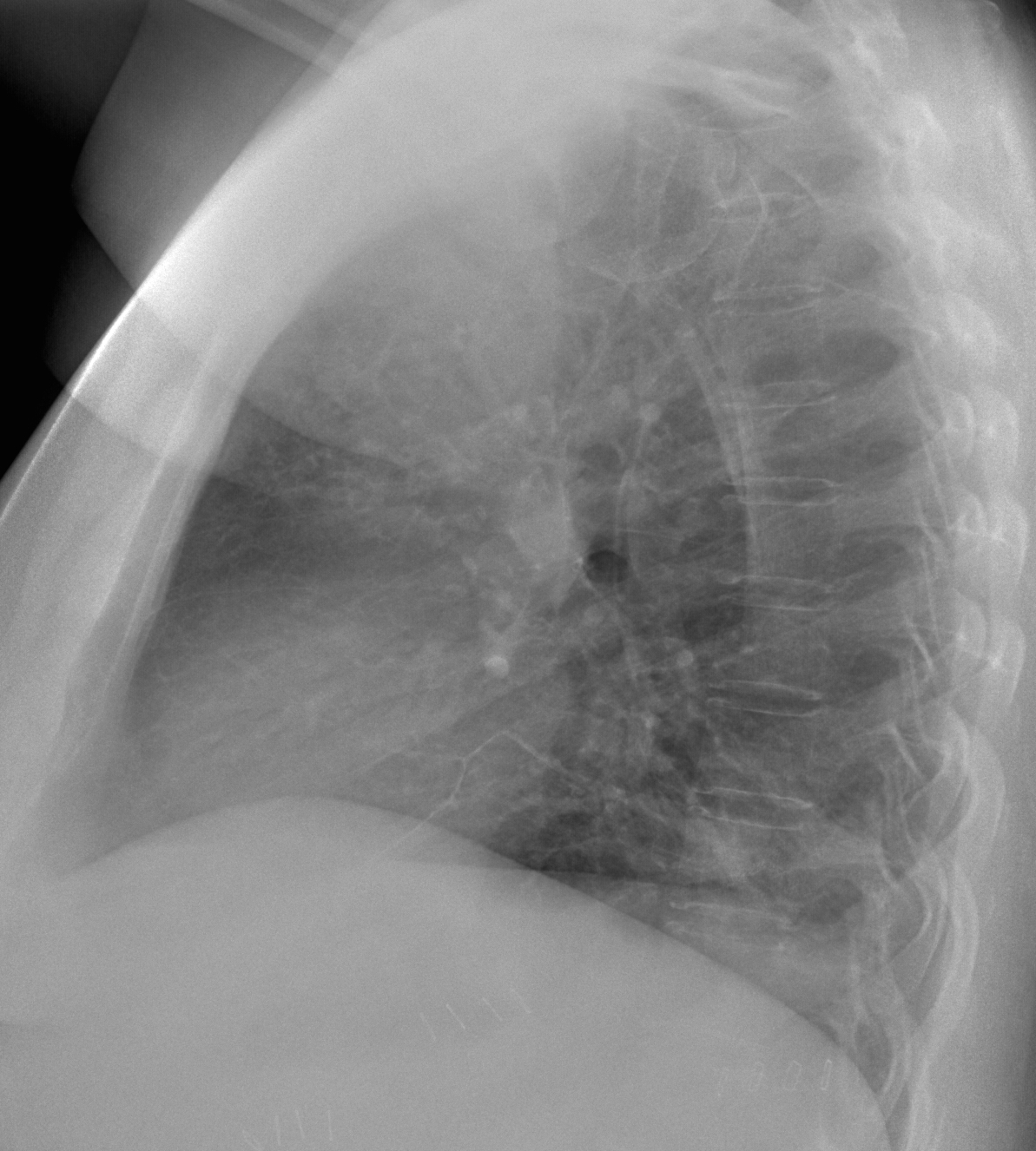
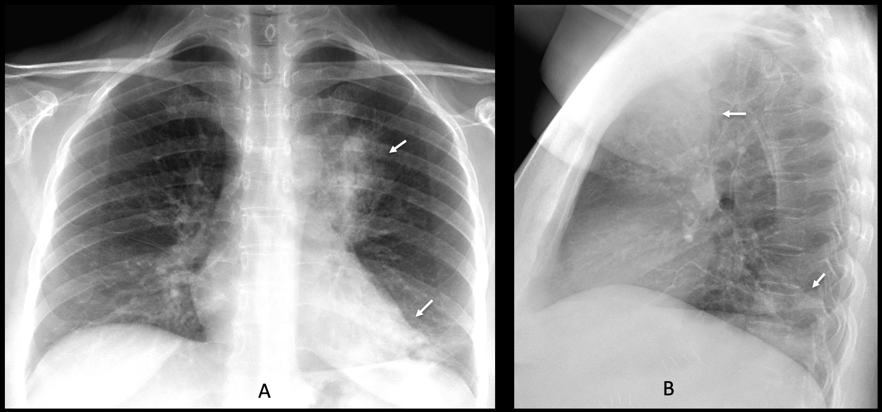
Today I am presenting the last case of the season. These images belong to a 47-year-old woman with a chronic cough and mild fever. The pulmonary infiltrate has not changed significantly in the last six months.
Check the images below, leave your thoughts in the comments section, and come back on Friday for the answer.
Findings: Chest radiographs show air-space disease in the LUL and LLL (A-B, arrows).
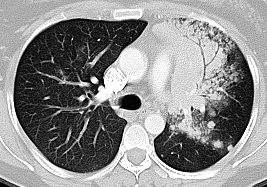
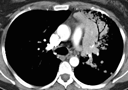
Enhanced axial CT confirms the LUL air-space disease, with an obvious air bronchogram (C-D, red arrows).
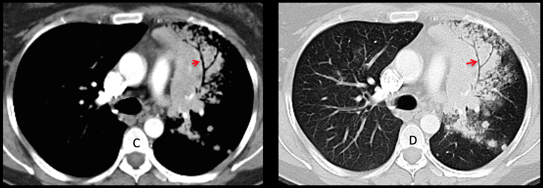
The bronchi are squeezed and scarcely branching. This appearance is highly suggestive of a malignant process that infiltrates the alveolar space, either adenocarcinoma or lymphoma (see Dr. Pepe’s Diploma Casebook Case #62). Biopsy confirmed the diagnosis of adenocarcinoma.
Control radiographs taken one year later show progression of the disease (E-F, arrows).
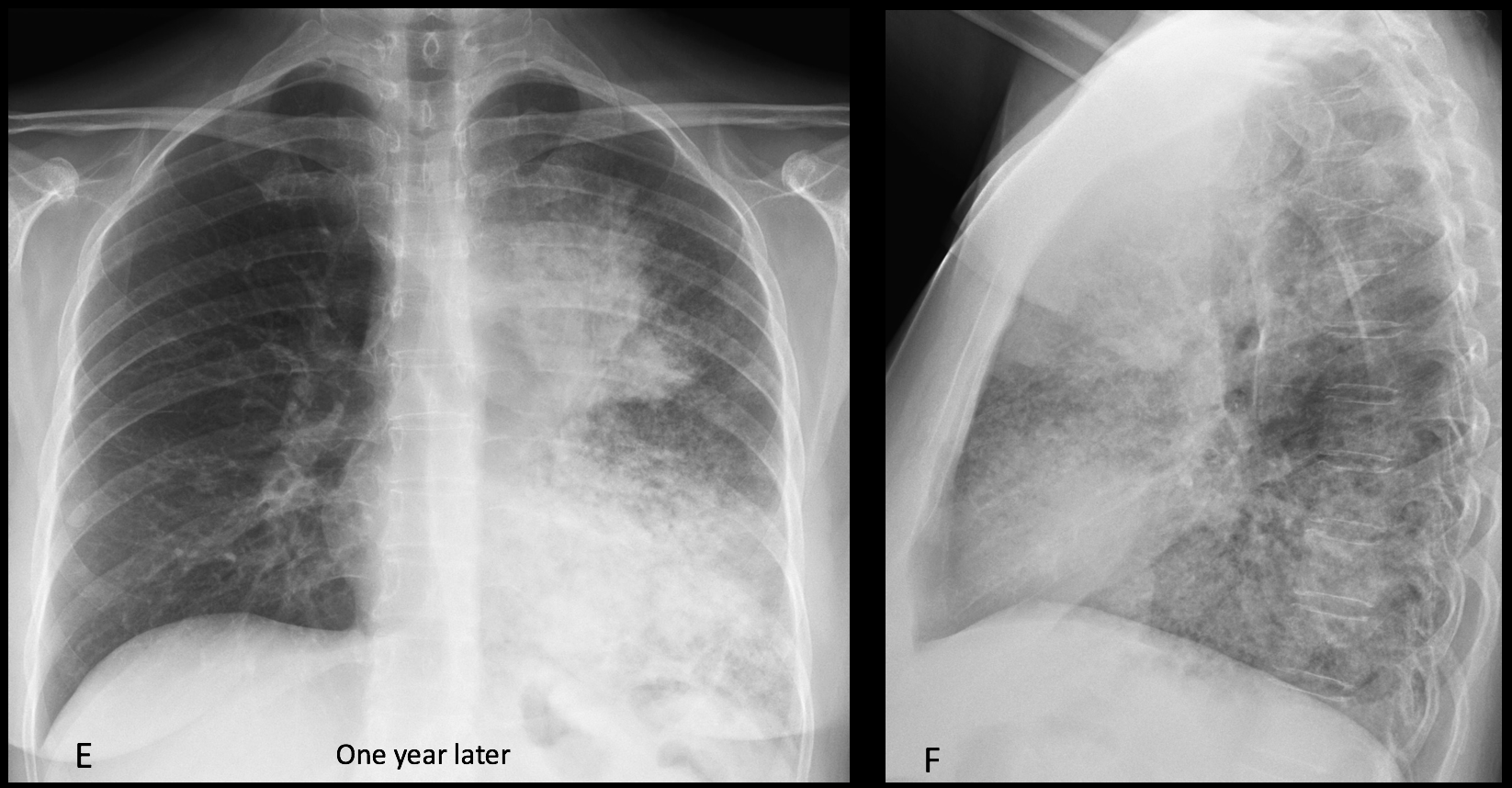
Final diagnosis: adenocarcinoma of the lung (alveolar-cell variant).
Congratulations to Amjad Ramzanali, who suggested the correct diagnosis.
Teaching point: remember that dilated bronchi are usually seen in inflammatory opacities, where the volume loss makes the bronchi shorter and wider. Narrowed bronchi, the so-called leafless tree appearance, occur in neoplastic infiltration of the alveolar space surrounding the bronchi.
This is the last case of the season. See you again in September.
Enjoy your vacation!