This case was provided by my good friend Alberto Villanueva, who is a fan of Real Madrid. Aside from that, he is normal.
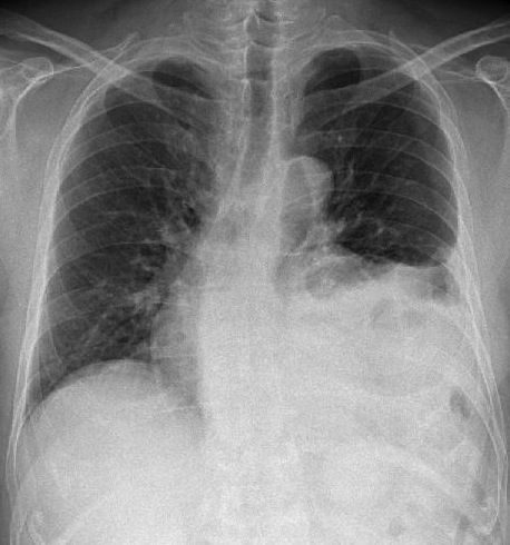
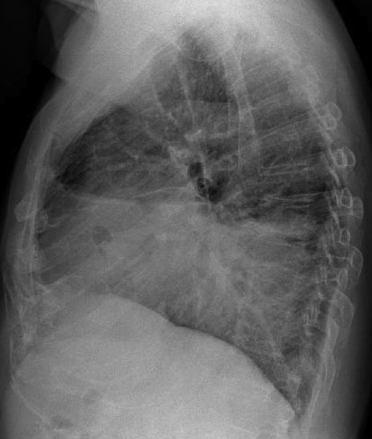
Preoperative chest in a 68-year-old man with a 6cm abdominal aortic aneurysm.
1. Leaking aneurysm
2. Fibrous tumor of pleura
3. Chronic empyema
4. None of the above
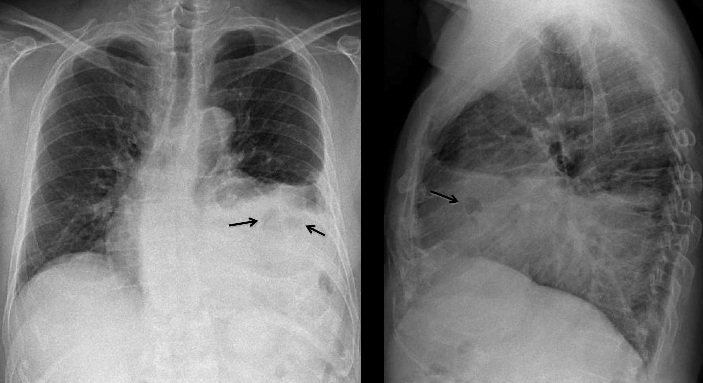
PA and lateral chest show a large opacity in the lower lung field, with an irregular upper border. There is blunting of the costophrenic angle and areas of air-density within the opacity (arrows), which resemble bowel loops.
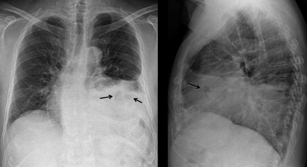
Once bowel loops are suspected, the choices are between hernia and diaphragmatic paralysis. An elevated diaphragm usually has a concave, well-defined border, whereas hernias have an irregular and flatter contour. If previous trauma is elicited, traumatic hernia should be ruled out. This patient had a severe accident fifteen years earlier and had been asymptomatic since.
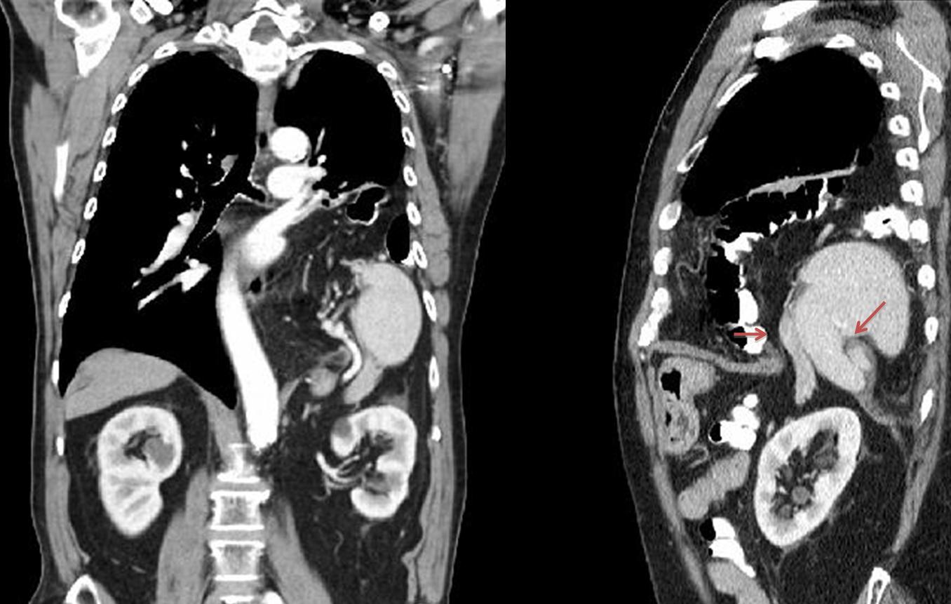
Sagittal and coronal CT show a rent in the diaphragm (arrows) with herniation of the spleen, colon and mesenteric fat.
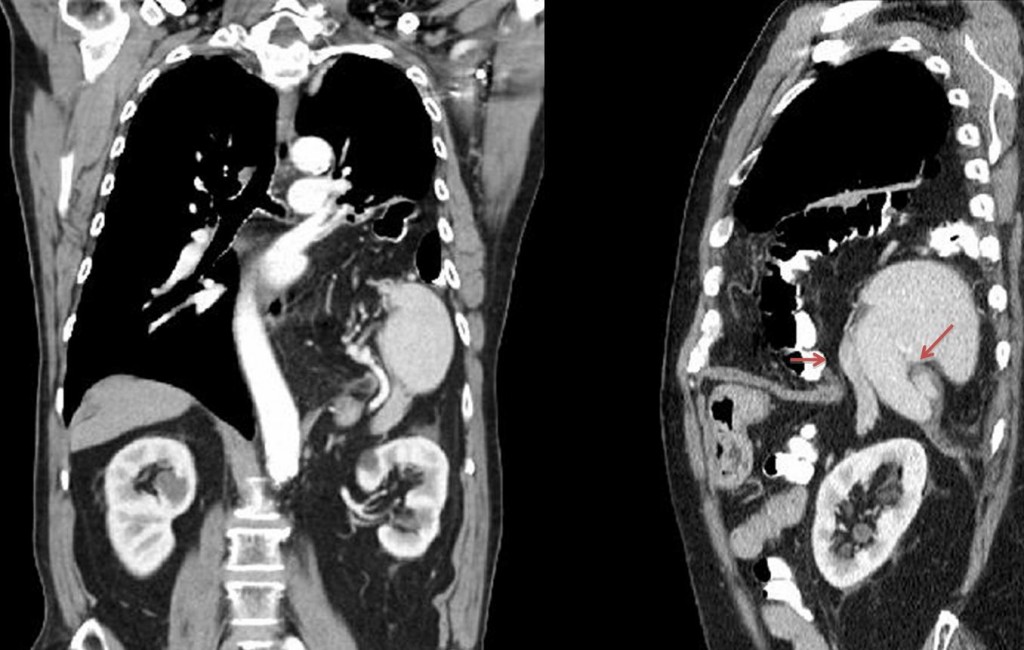
Final diagnosis: traumatic diaphragmatic hernia.
Muppet is on vacation in Menorca and congratulates the faithful few who participated and answered correctly.
Teaching point: the diaphragm is a frontier organ. Any density in the lower lobes and in contact with the diaphragm may arise from below.