Dr. Pepe’s Diploma Casebook: Case 39 – SOLVED!
Presenting mammograms of a 45 y.o. woman with a palpable abnormality in the left breast.
Diagnosis:
1. Carcinoma
2. Fibroadenoma
3. Intrammamary lymph node
4. All of the above
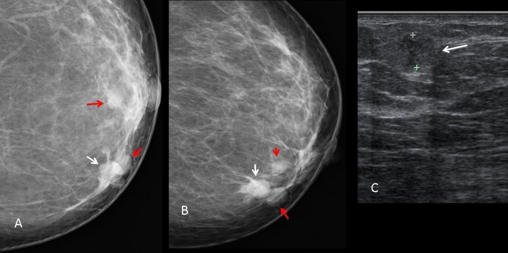
Findings: There is a nodule in the lower inner quadrant of the left breast (A, B arrows) with a partially ill-defined border. Two other nodules of lower density are also seen (red arrows). The main nodule was reported as BIRADS 4 B. Ultrasound demonstrates an irregular, solid nodule (C, arrow) with two nearby cysts. Core biopsy of the solid nodule was performed.
Final diagnosis: infiltrating ductal carcinoma.
According to Dr. D.B. Kopans, carcinoma is the only important disease of the breast.
The major role of mammography is early detection of breast cancer in asymptomatic women. Together with ultrasound it is highly useful in women with signs or symptoms of breast disease. The approach to imaging interpretation should emphasize management of the abnormal finding, rather than the specific diagnosis.
Discovery of a nodule in a woman older than 35 should always alert to the possibility of malignancy, especially if the nodule shows poorly-defined borders, although 10%-20% of well-circumscribed nodules are malignant. Mammography should then be complemented with ultrasound, which will add valuable information and will exclude simple mammary cysts. A solid nodule with irregular margins and poor sound transmission should undergo core biopsy (Fig. 2).
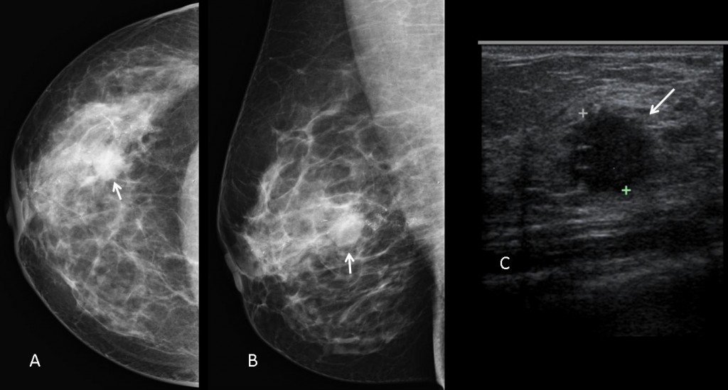
Fig. 2 (above): 36-year-old woman with a breast nodule showing poorly-defined borders (A,B arrow). Microcalcifications are seen in the surrounding parenchyma. Ultrasound shows a solid hypoechoic nodule with irregular margins (C, arrow). Biopsy: infiltrating ductal carcinoma.
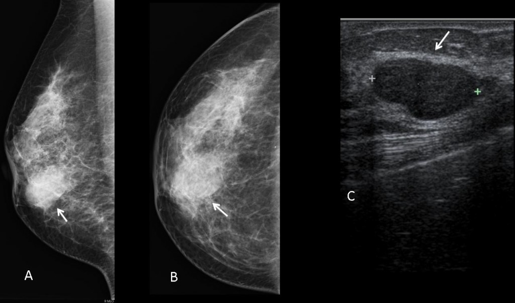
Fig. 3 (above): fibroadenoma of the breast. Mammograms show an ovoid nodule (A,B arrow) with ill-defined borders. When a nodule is surrounded by dense fibroglandular tissue it is difficult to differentiate between an invasive margin and one that is obscured by normal breast tissue. Ultrasound shows the typical appearance of fibroadenoma with homogeneous echogenicity, well-defined borders, and good sound transmission (C, arrow). Nevertheless, any solid nodule in a woman older than 40, regardless of its imaging appearance, should always undergo core biopsy if there is any doubt of malignancy.
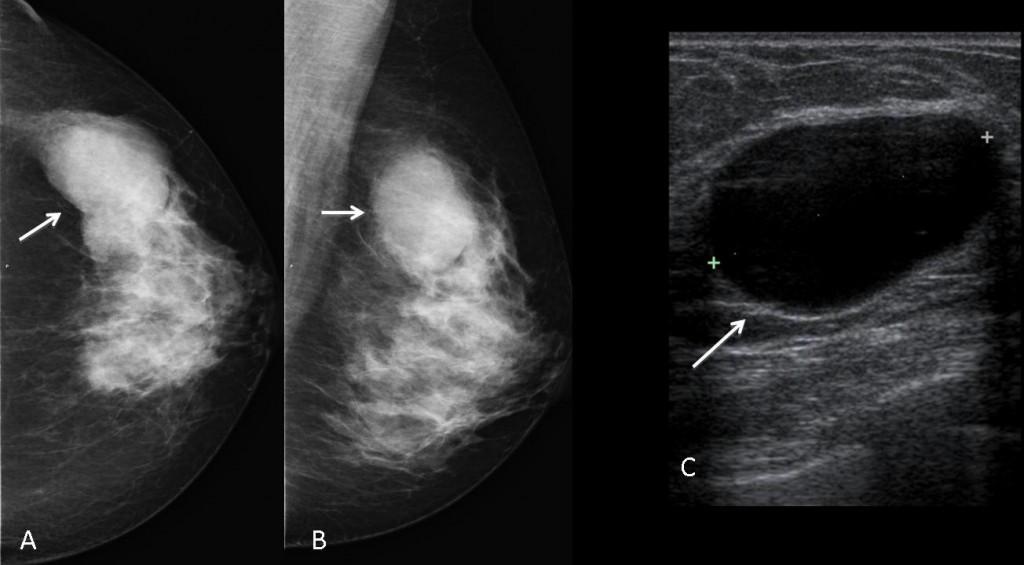
Fig. 4 (above): simple breast cyst in a 48-year-old woman. Mammography shows an ovoid nodule (A,B, arrows) that is anechoic on ultrasound study (C, arrows).
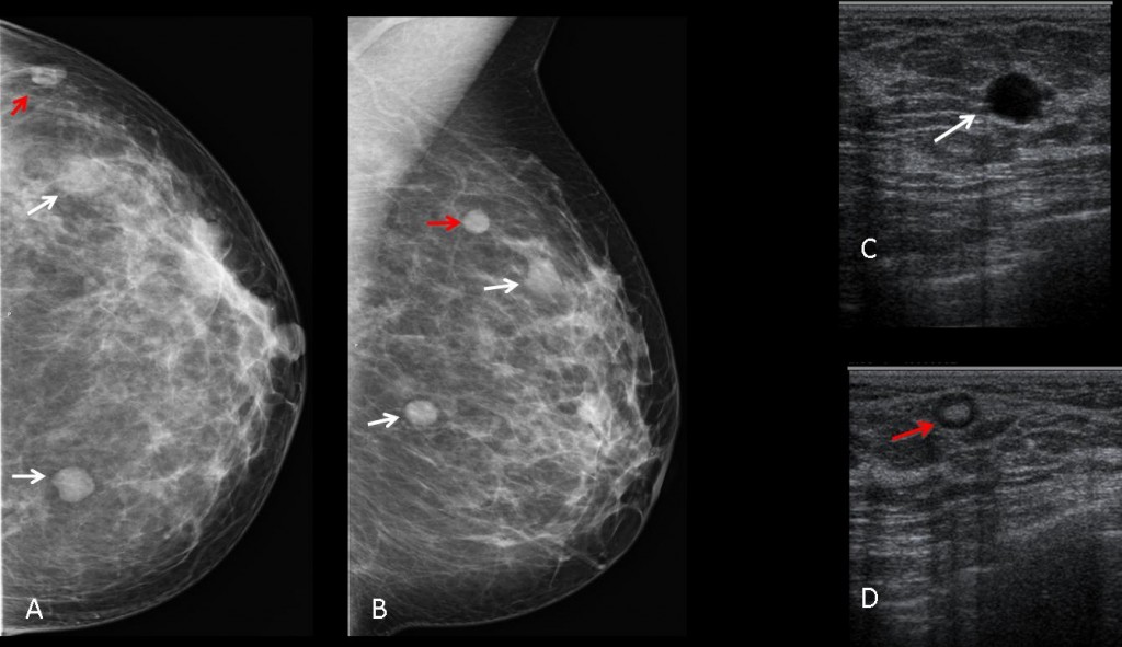
Fig. 5 (above): 38-year-old woman with three mammary nodules (A,B arrows). Ultrasound shows that two of them are cysts (C, arrow). The third one, located in the upper outer quadrant (A,B, red arrow) has the typical appearance of a lymph node, with an echogenic fatty center (D, arrow).
- The most common nodular lesions of the breast are cysts, carcinomas, fibroadenomas and lymph nodes.
- Ultrasound should be performed in all nodular lesions to exclude cysts.
- In solid nodular lesions, malignancy should be excluded. When in doubt, core biopsy should be performed
Recommended reading: DB Kopans, Breast Imaging, 3rd Ed. Chapter 13
Case prepared by Elena Rabanal, MD




. 2. Fibroadenoma
cancer
2. Fibroadenoma
4.
4. All of the above
4.All of the above
Option 4
ectasia duttale.