Dr. Pepe’s Diploma Casebook: Case 51 – SOLVED!

Dear Friends,
I am back with radiographs of a 63-year-old woman with malaise and low-grade fever. Check the images below and leave me your thoughts and diagnosis in the comments section. Come back on Friday for the answer.
Diagnosis:
1. Carcinoma of the lung
2. Pneumonia
3. Thymoma
4. None of the above
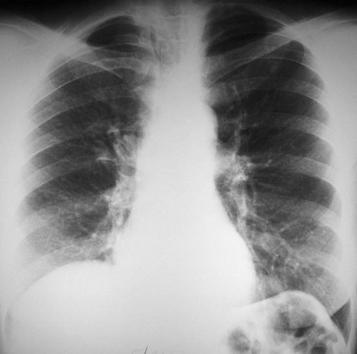
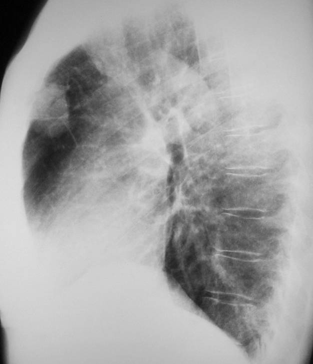
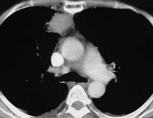
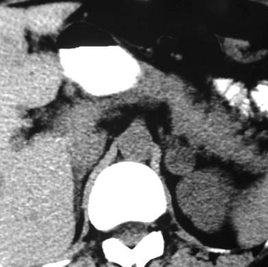
Findings: the PA view of chest is unremarkable. The lateral view shows a nodular shadow in the anterior clear space (A, arrow). Its borders are ill-defined, both in the radiograph and the CT image (B, arrow), indicating an intrapulmonary lesion and excluding thymoma. There is a non-enhancing 2cm nodule in the left adrenal gland (C, arrow).
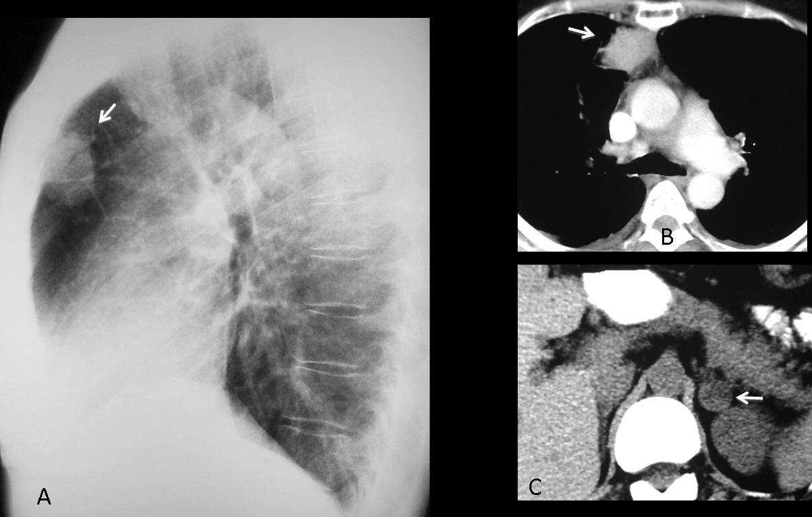
Fig. 1
Because the clinical findings were consistent with acute infection, it was decided to treat the patient with antibiotics. Radiographs and CT taken two weeks later show that the mass had markedly decreased in size.
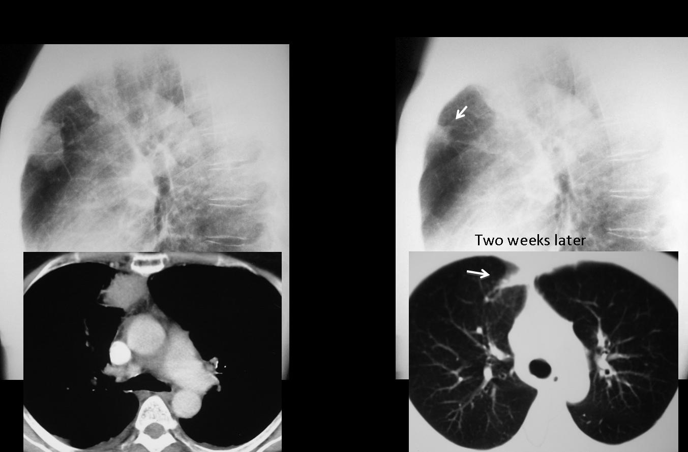
Fig. 2
Final diagnosis: Round pneumonia. Incidental adrenal adenoma
In adults, most lung nodules represent carcinomas or granulomas. In this presentation I want to discuss innocuous nodular lung lesions in which symptoms or radiography findings suggest the correct diagnosis, confirmed with CT. Some of them are transient (round pneumonia, pulmonary haematoma) and others are stable, remaining unchanged in serial studies.
The first such lesion is the so-called round pneumonia, which appears as a rounded lesion in the chest radiograph. Round pneumonias usually occur in childhood, but they are also seen in adults. They are due to incomplete development of collateral airways, limiting spread of an infection and resulting in a rounded infiltrate.
In the appropriate clinical setting (acute fever, cough), round pneumonia should be suspected when a rounded lung lesion is seen in an adult. Before CT is performed, a follow-up film should be taken to confirm or exclude this possibility (Fig. 3).
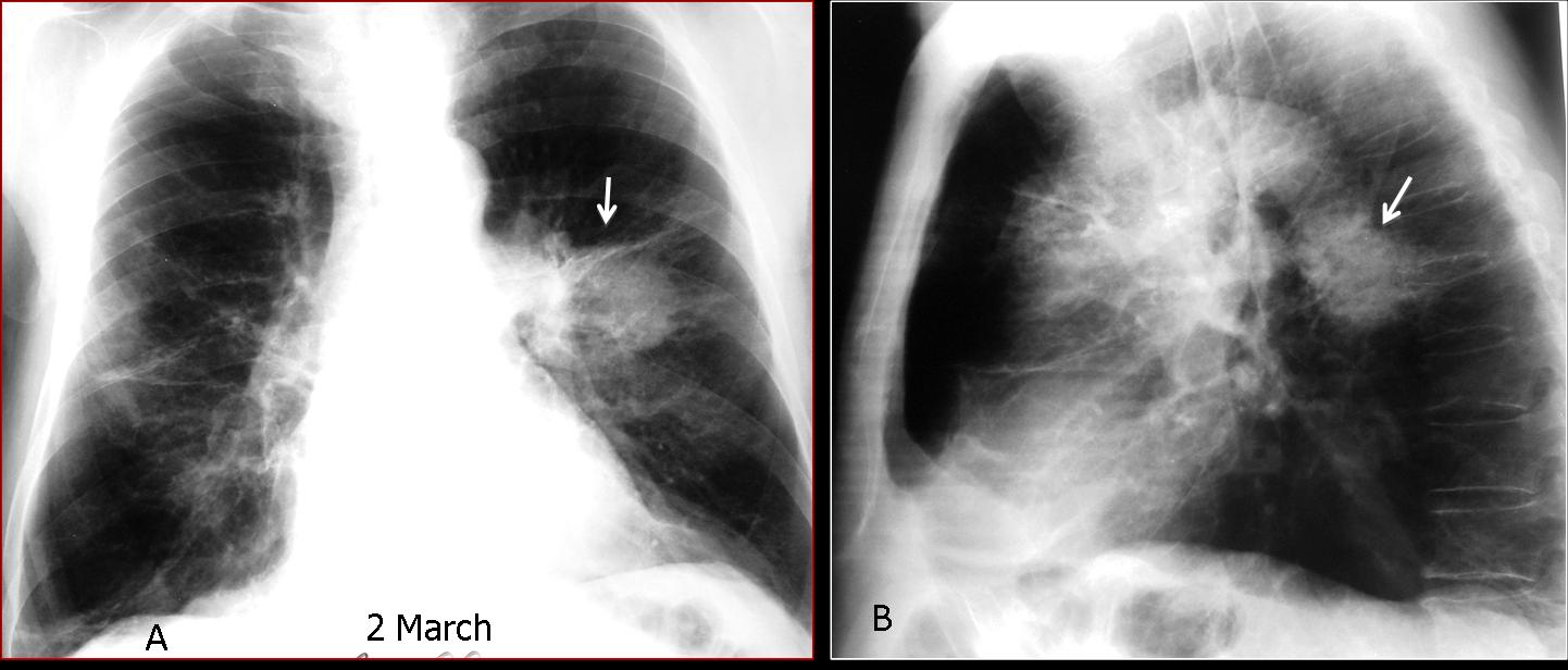
Fig. 3
Fig. 3 (above): 68-year-old smoker with cough and high fever. Chest radiographs show a round lesion in the left lung suspected to be carcinoma (A, B, arrows). Because of the fever, antibiotic treatment was started. Seven days later, the round lesion has disappeared and a linear scar remains (C, arrow). Diagnosis: round pneumonia.
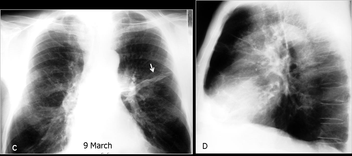
Fig. 4
Pulmonary haematoma is a transient nodular lesion secondary to localised parenchymal haemorrhage due to a traumatic injury or coagulation disorder. In the first scenario, blunt trauma causes a laceration in the lung parenchyma that fills with blood. Spontaneous haematomas are usually related to anticoagulation therapy. In both cases they present as a relatively high attenuation nodule on CT that decreases in size over time. The clue to suspecting the diagnosis is the background of trauma or the blood dyscrasia. When haematoma is suspected, follow-up films confirm progressive shrinkage of the nodule and its eventual disappearance (Figs. 5, 6 and 7).
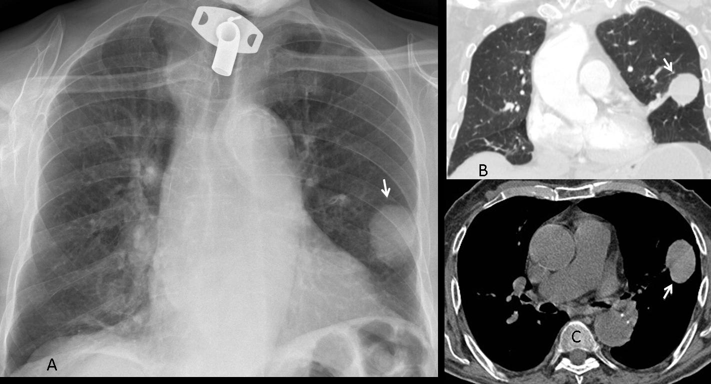
Fig. 5
Fig. 5a (above): 75-year-old man with acute pulmonary embolism treated with anticoagulants. During his stay at the hospital, a round nodule appeared in the left lung (A,B arrows), showing high density in the CT study (C, arrow). It was interpreted as a spontaneous haematoma related to the treatment. The nodule progressively decreased in size and disappeared three months later, leaving a linear scar (Fig. 6 A,B, arrows).
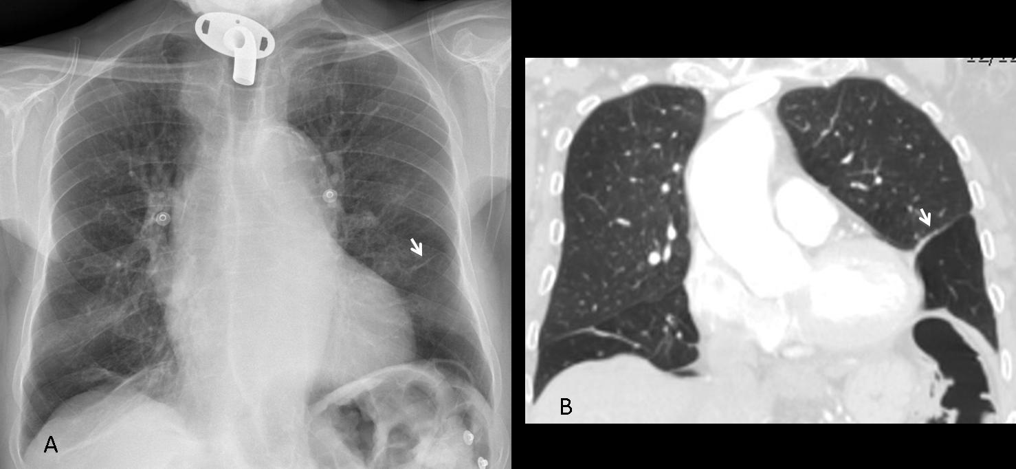
Fig. 6
Fig. 7 (below): 27-year-old man following a motorcycle accident. CT shows several pulmonary haematomas (A,B, white arrows), surrounded by areas of pulmonary contusion (B, red arrows). One month later, the RLL hematoma has disappeared, and the others have decreased markedly in size (C,D, arrows).
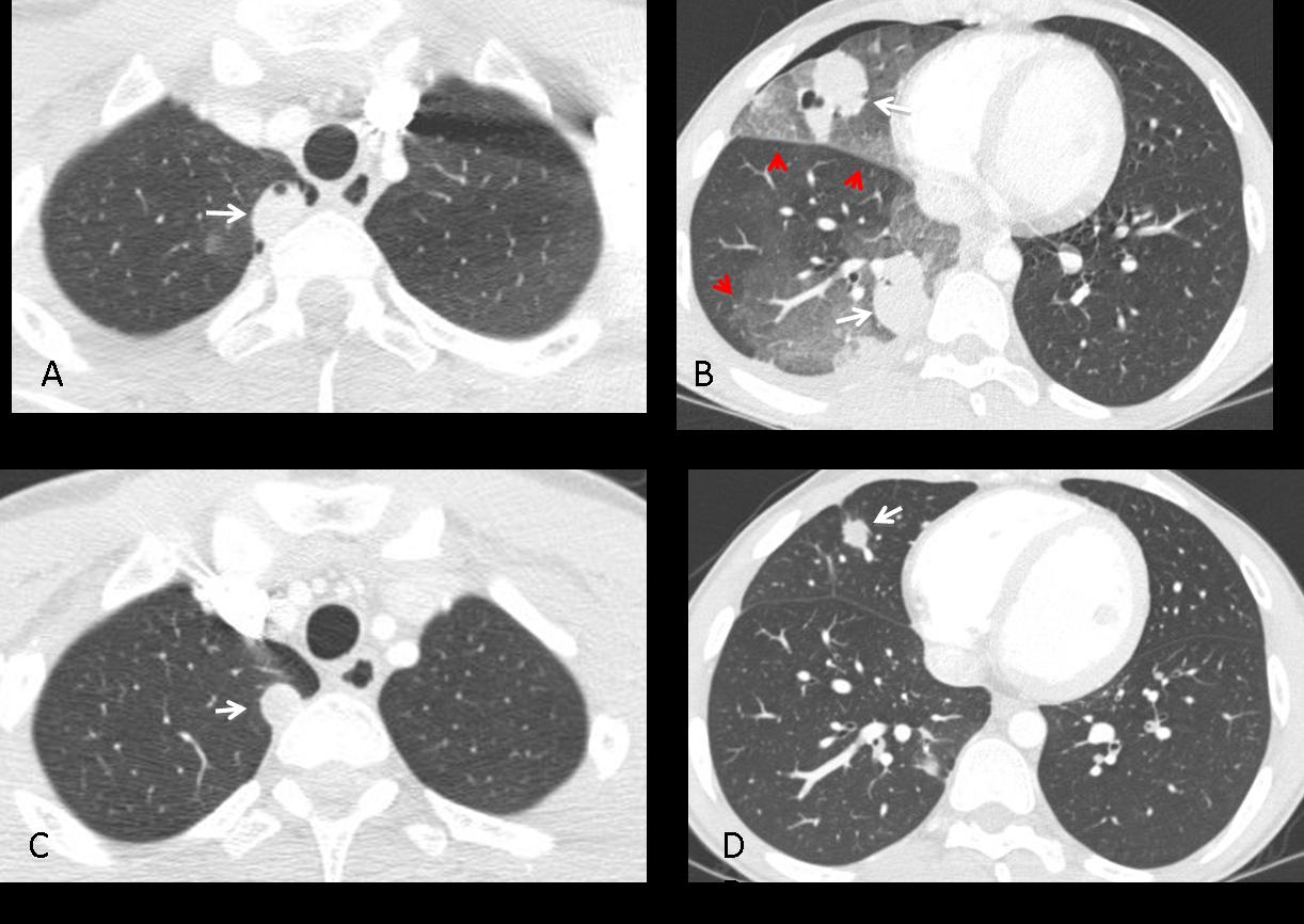
Fig. 7
Rounded atelectasis is not an uncommon cause of a stable nodular lesion. It occurs secondary to spiral folding of the lung parenchyma when fixed by thickened pleura. The consequence is a peripheral rounded opacity which should not be confused with a true nodule. It is usually asymptomatic and associated with asbestos-related disease. The imaging appearance of rounded atelectasis is very characteristic. The typical features include:
1. Peripheral lung nodule
2. Pleural thickening
3. Loss of volume of the affected lobe
4. Curving of vessels and bronchi on CT
These features are well demonstrated in Fig. 8.
TIP: Since rounded atelectasis occurs as a consequence of pleural disease, a lack of pleural thickening should lead us away from this diagnosis and suggest other possibilities.
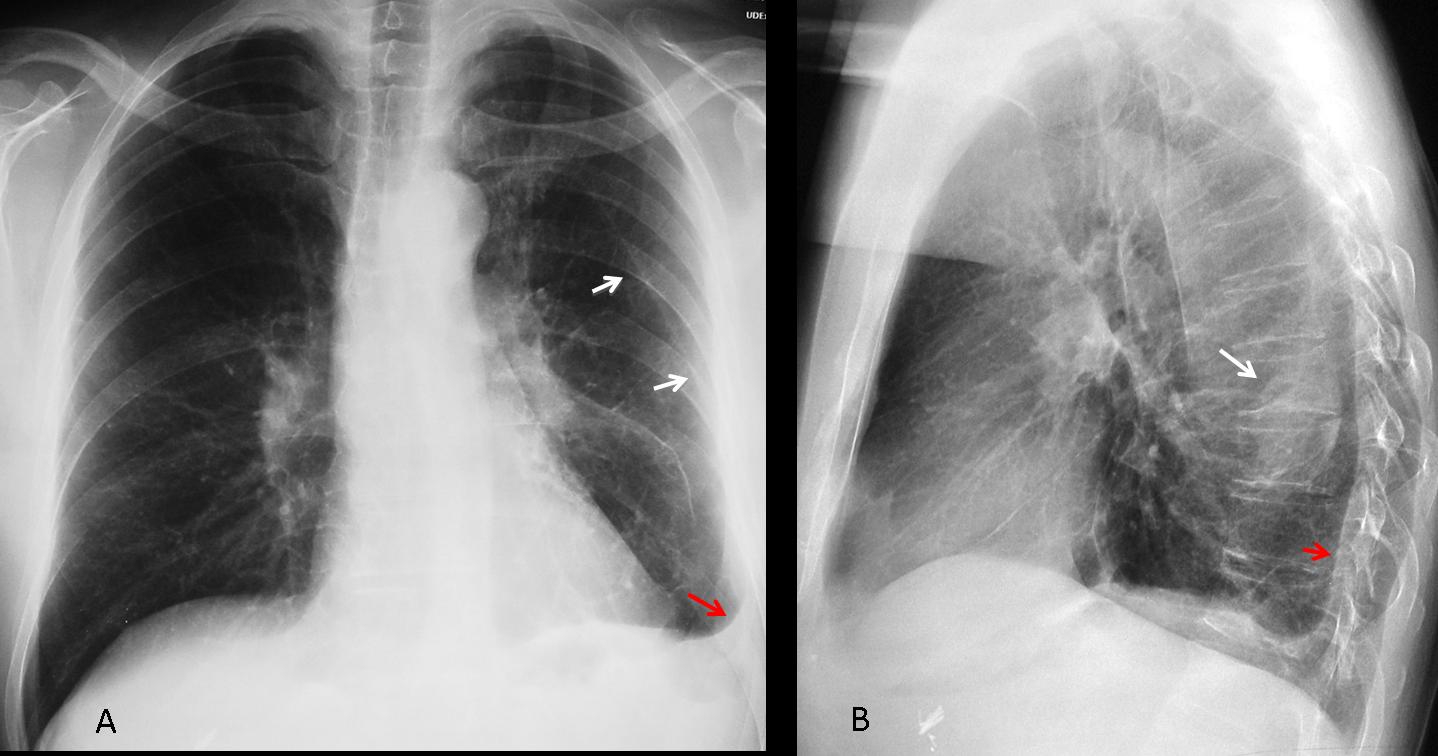
Fig. 8
Fig. 8 (above): asymptomatic 49-year-old man with rounded atelectasis. Notice the peripheral nodular component (B, white arrow), inner displacement of left major fissure indicating loss of LLL volume (A, white arrows), and the pleural disease (A, B, red arrows).
CT shows pathognomonic signs: curving of the vessels into the atelectatic nodule (Fig. 9 A,B, white arrows), which also has an air bronchogram in the proximal portion (B, blue arrows). Note the pleural thickening (A,B, red arrows).
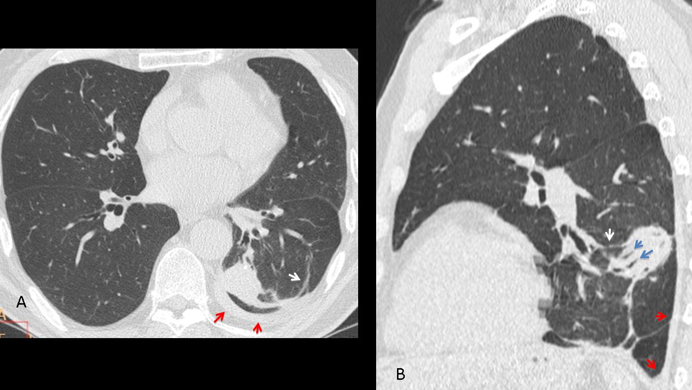
Fig. 9
At times, rounded atelectasis may be difficult to diagnose in the plain film; the typical features are discovered when performing CT to study an indeterminant nodule (Fig. 10)
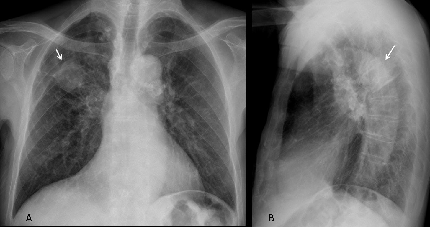
Fig. 10
Fig. 10 (above): rounded atelectasis in a 63-year-old man with an indeterminate nodule in the chest film (A, B arrows). The minor fissure is elevated, indicating RUL volume loss.
CT images show that the nodule represents rounded atelectasis. Note the peripheral localisation with associated pleural thickening (Fig. 11 A,B, red arrows), the curving vessels (A,B white arrows) and the air bronchogram in the proximal portion (B, blue arrows).
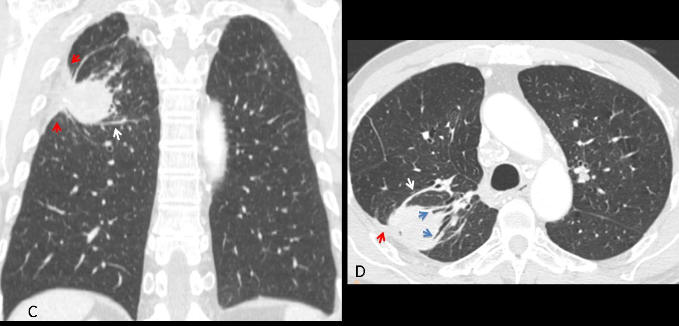
Fig. 11
Time for a test. Below is the PA radiograph of a 47-year-old asymptomatic man, in whom an RUL nodule was found. What would your diagnosis be?
1. Granuloma
2. Hamartoma
3. Mucus plug
4. None of the above
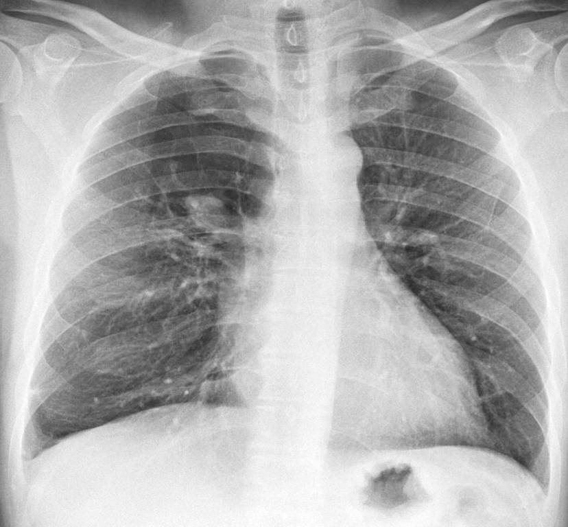
Fig. 12
Findings: PA radiograph shows a right parahilar nodule (A, arrow). Most of the right upper lung is hyperlucent compared to the left side. The combination of a central nodule and peripheral hyperlucency is suspect of mucus plugging with distal overinflation. Coronal CT shows the mucus plug (B, arrow) and the lack of bronchi in the right upper lung. Diagnosis: mucus plug in congenital bronchial atresia.
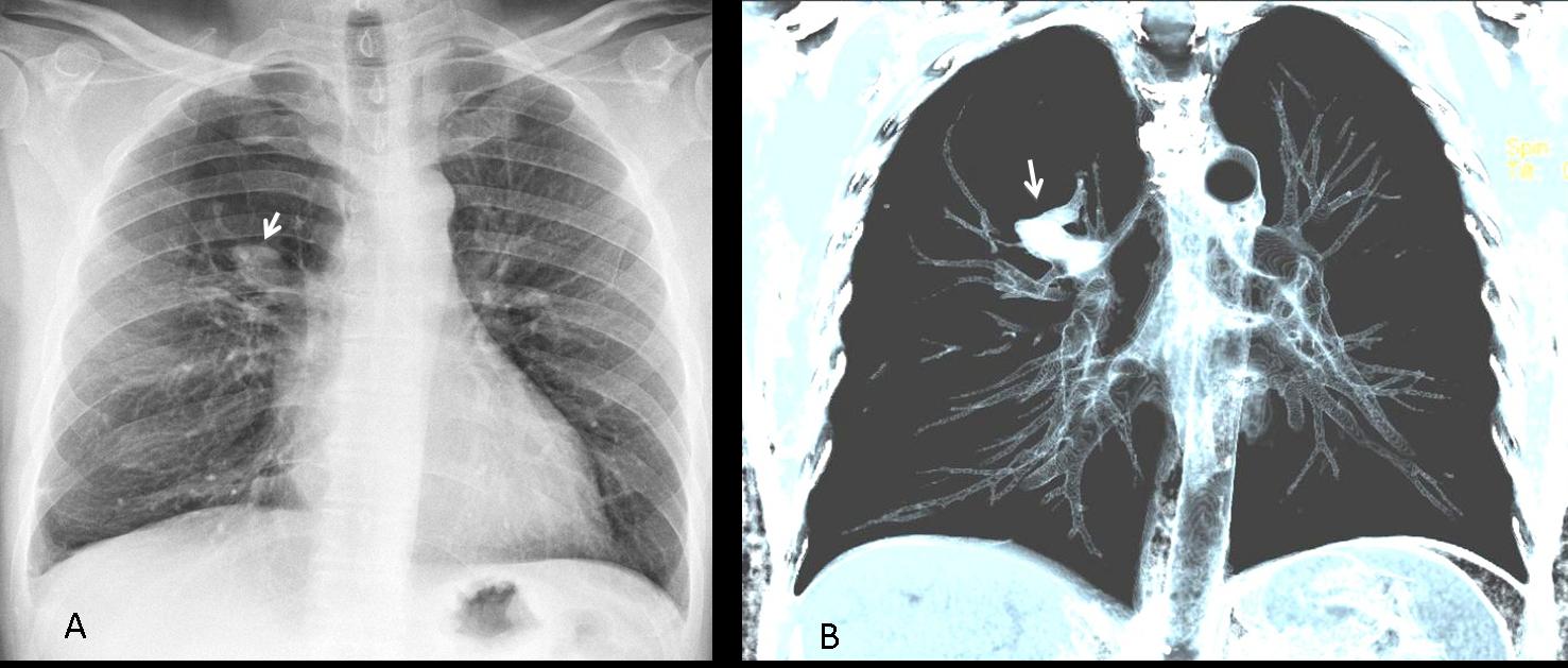
Fig. 13
Mucus plugs occur when obstruction of a segmental bronchus allows the mucus to accumulate distally. They usually manifest as tubular opacities which, when seen end-on, may simulate a nodule. Mucus plugs can be seen in congenital and acquired conditions. In my experience, the hyperlucency associated with congenital bronchial atresia facilitates identification of the plug in the chest radiograph. CT easily confirms the diagnosis (Fig. 14).
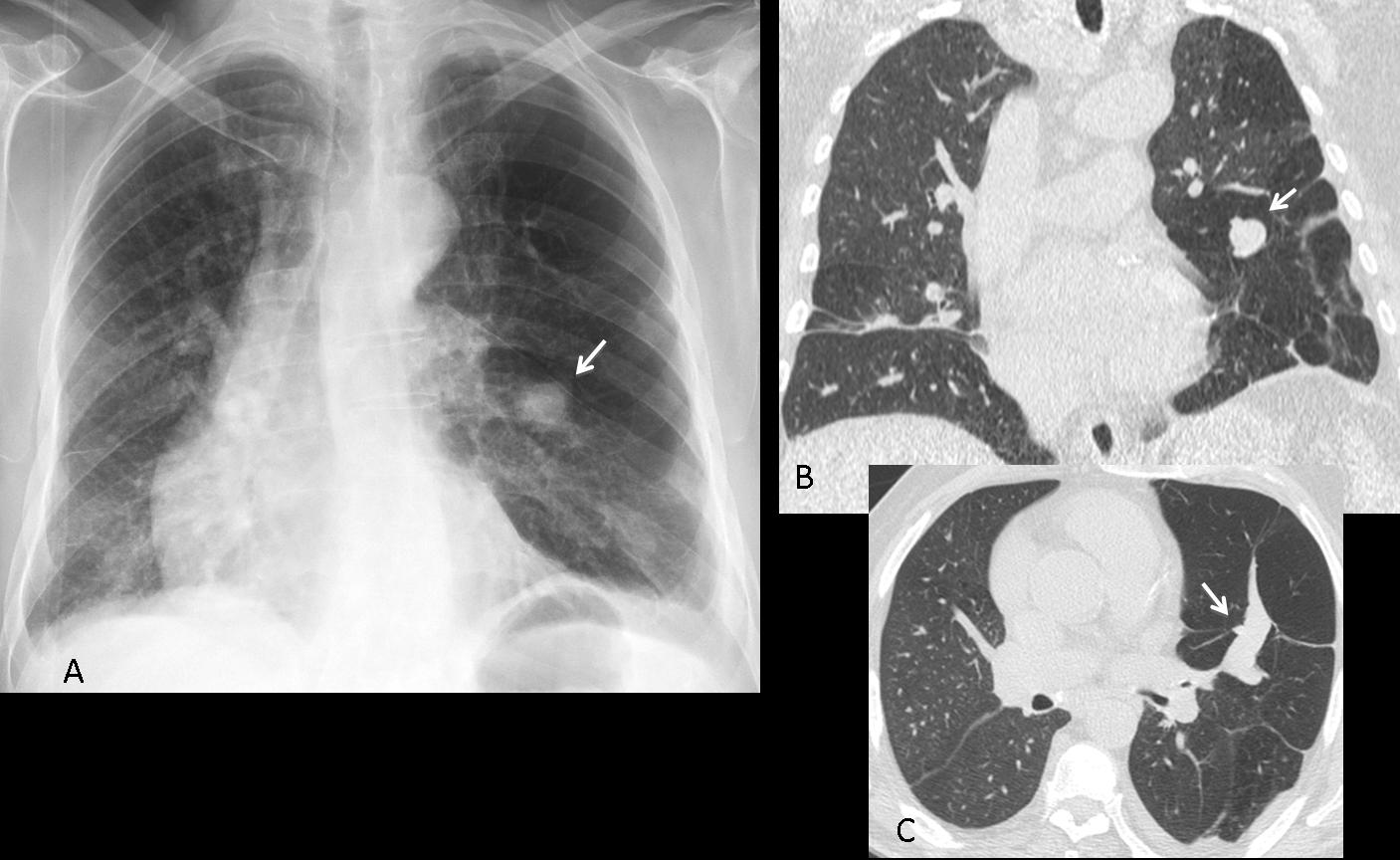
Fig. 14
Fig. 14 (above): 64-year-old man with cough and moderate dyspnea. PA chest film shows left lung hyperlucency with a nodule in the middle field (A, arrow). Coronal CT confirms the nodule (B, arrow) and hyperlucency of lower lung. Axial CT depicts the typical appearance of a mucus plug (C, arrow). Diagnosis: congenital bronchial atresia.

Follow Dr. Pepe’s advice
Follow Dr. Pepe’s advice:
- Not all nodular lung lesions represent carcinomas or granulomas.
- Round pneumonias and hematomas present as transient nodular lesions, which are easily suspected based on the presenting symptoms and confirmed with follow-up films.
- Rounded atelectases are peripheral nodules associated with pleural disease and loss of volume of the affected lobe. CT appearance is pathognomonic.
- When seen end-on, mucous plugs occasionally simulate nodular lesions. The association with hyperlucency suggests bronchial atresia. CT is diagnostic.


chest, CT, diagnosis, EDiR, European Diploma in Radiology, radiography