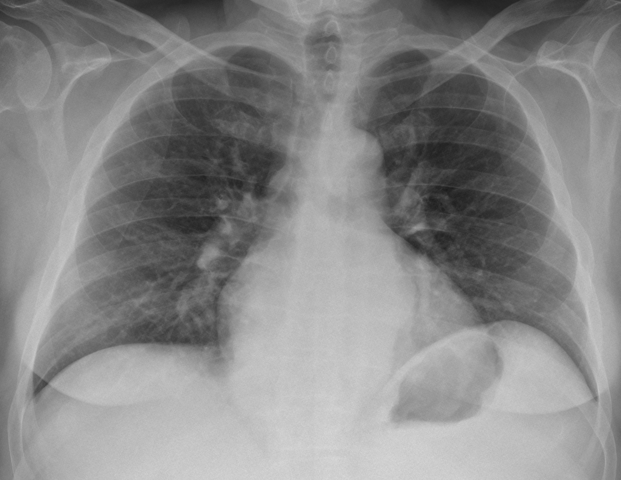
Muppet and I are very happy to have reached one hundred cases. We hope you enjoyed them as much as we did. Radiographs of this case belong to a 52-year-old man with vague chest complaints. He was operated on for testicular tumour fifteen years earlier.
Check the images below, leave your thoughts and diagnosis in the comments section, and come back on Friday to find out the answer.
1. Duplication cyst
2. Lymphangioma
3. Metastasis from testicular tumour
4. None of the above
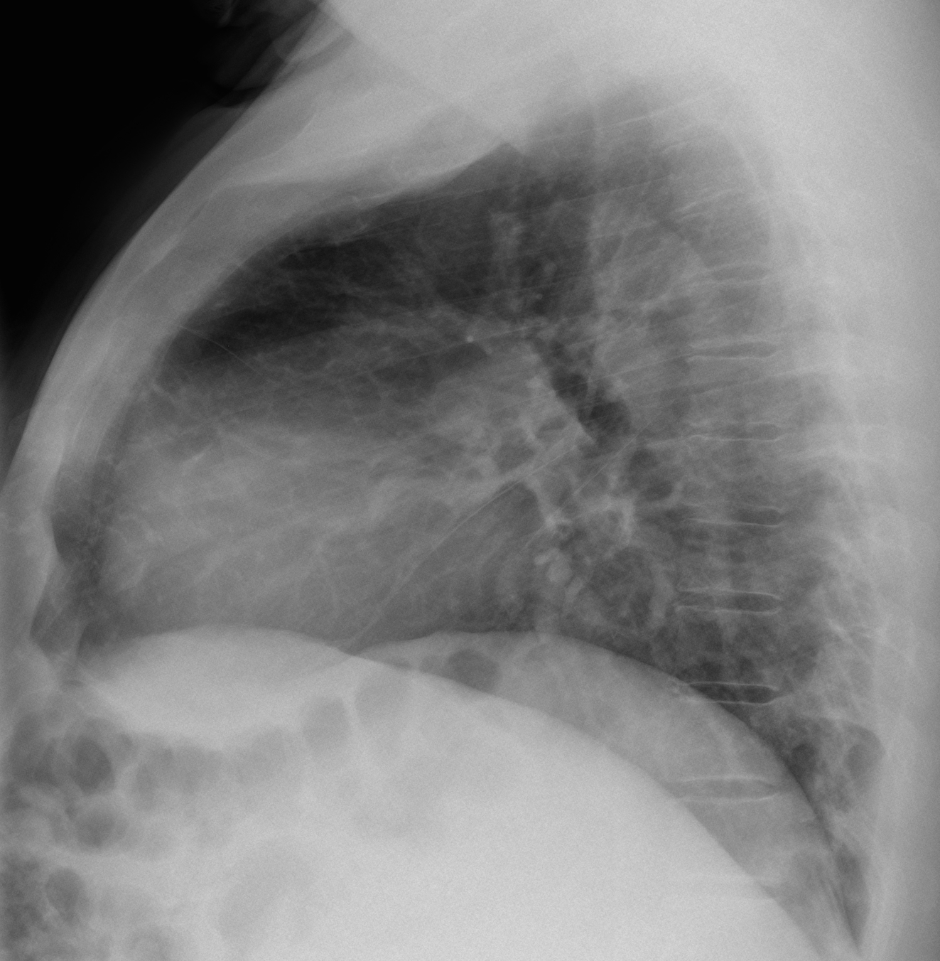
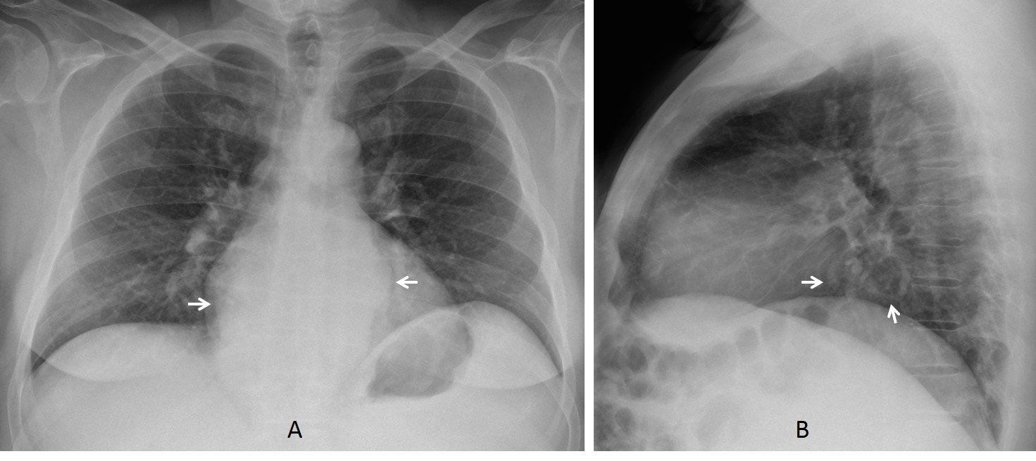
Findings: PA radiograph shows widening of lower mediastinal lines, which are slightly undulated (A, arrows). There is increased density in the left upper quadrant of the abdomen and the lateral wall of the stomach is indented, suggesting splenomegaly. On the lateral view there is increased opacity of the middle lower mediastinum, with a suggestion of tubular structures (B, arrows).
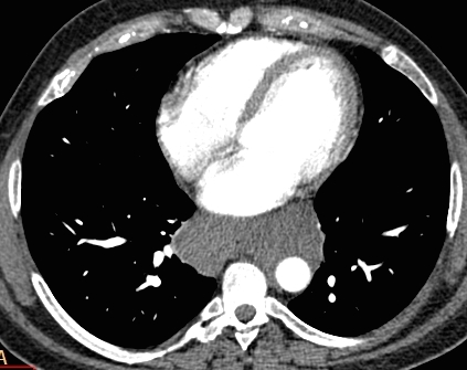
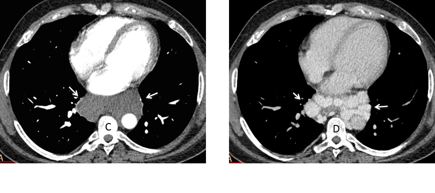
Enhanced axial CT (arterial phase) shows a non-enhancing mass in the middle mediastinum (C, arrows). Venous phase shows multiple enhancing veins within the mass (D, arrows).
Axial and coronal CT (E, F) show marked liver irregularity and splenomegaly. Note how the enlarged spleen impinges on the stomach (F, arrow).
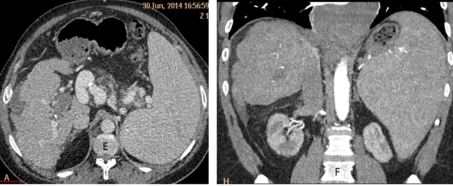
Final diagnosis: paraesophageal varices secondary to liver cirrhosis
I am showing this case as a reminder that vascular structures should always be considered when facing a mediastinal mass. A similar case was presented three years ago (Case #8). The same teaching point applies today.
Teaching point: the mediastinum is composed mainly of vascular structures. When a mediastinal abnormality is present, always rule out a vascular origin (arterial or venous).