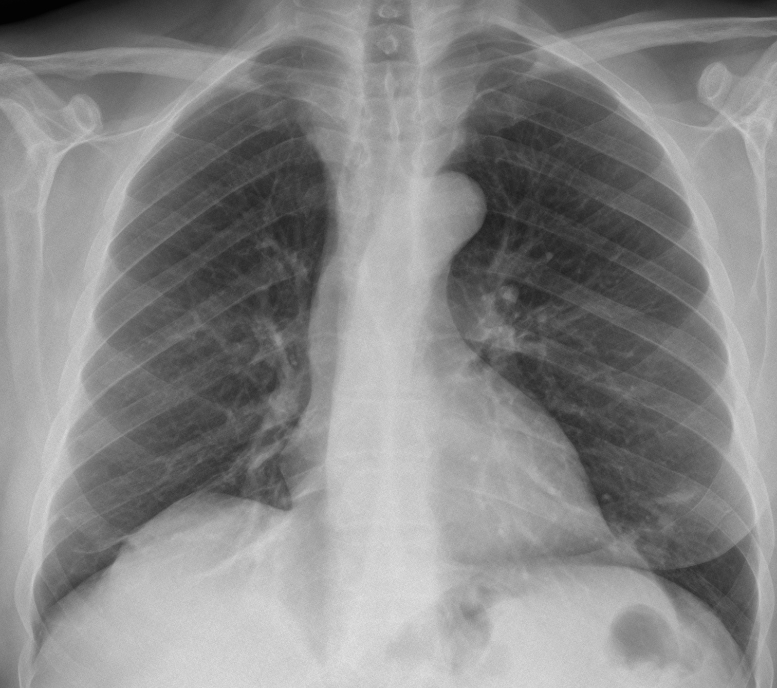
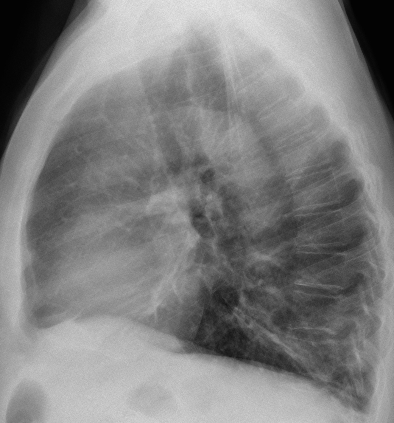
Today we are showing radiographs of a 61-year-old man who has had several pulmonary infections during recent years.
What do you see? Leave your thoughts in the comments section and come back for the answer on Friday.
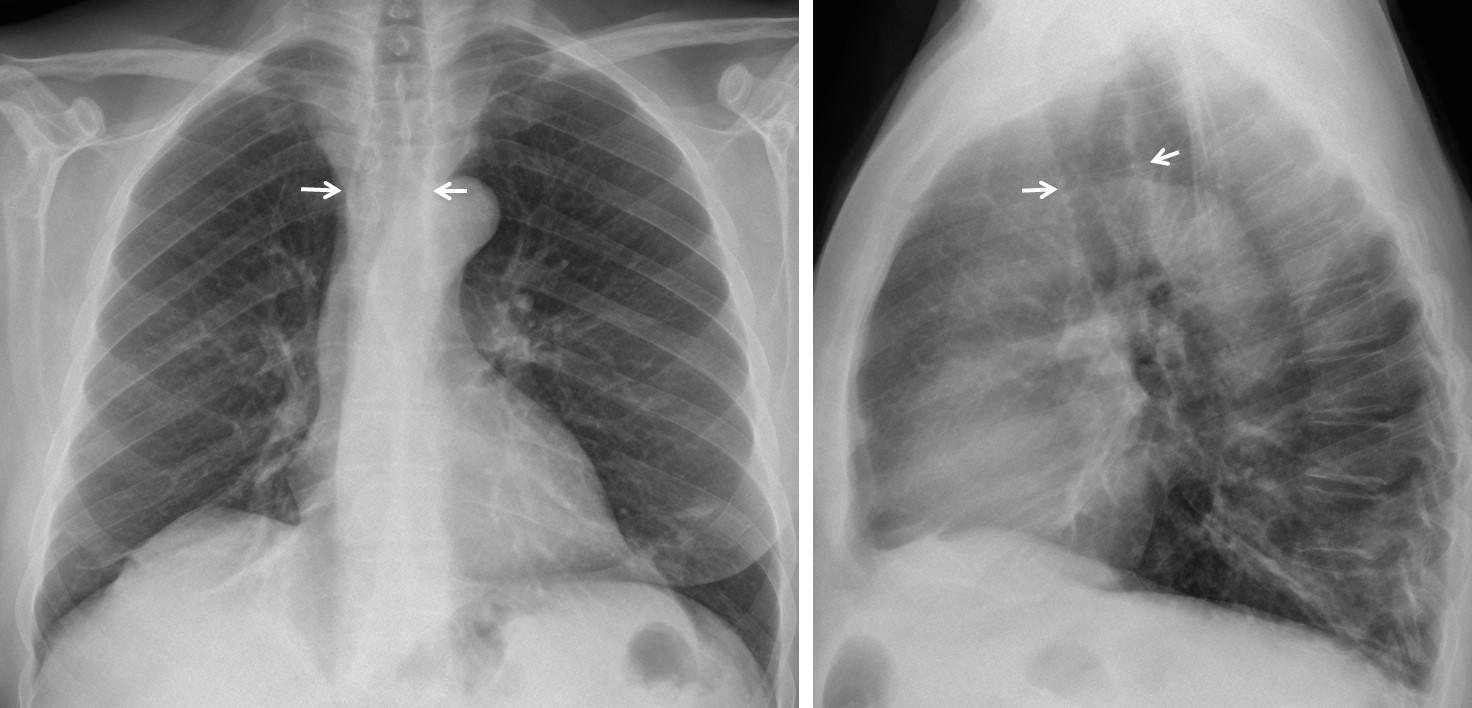
Findings: Chest radiographs show a marked increase of the tracheal diameter, both in PA and lateral views (arrows). Lineal infiltrates in both bases are seen, probably secondary to previous infections.
Unenhanced coronal and sagittal CT confirm the findings, showing a corrugated appearance of the tracheal wall and dilatation of the main bronchi. These findings are very characteristic of tracheobronchomegaly.
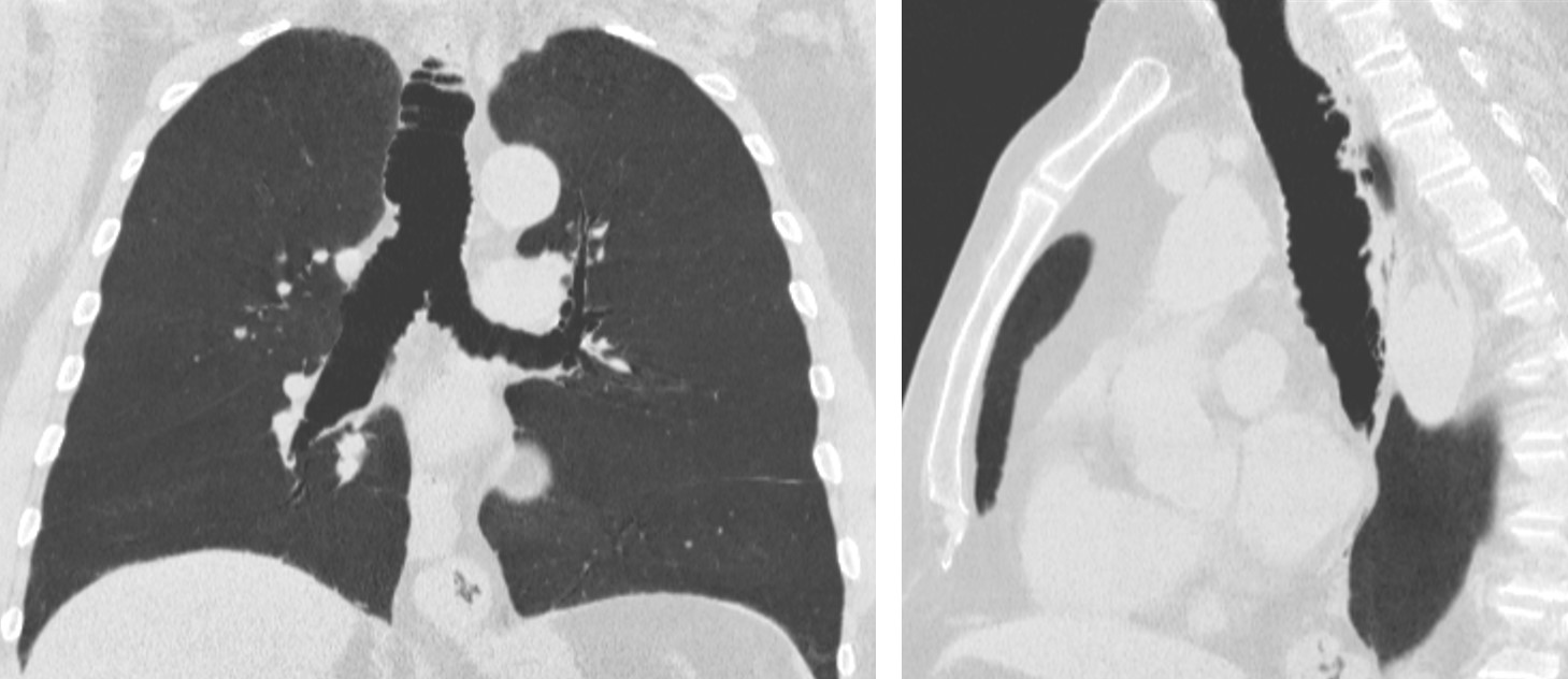
Final diagnosis: tracheobronchomegaly (Mounier-Kuhn syndrome)
Contratulations to Stanko, who was the first to suggest the diagnosis.
Teaching point: remember to look at the trachea when examining a chest film. Important findings can be discovered. Although tracheobronchomegaly is an unusual disease, the present case was seen in October of this year and the diagnosis was suggested in the plain film.