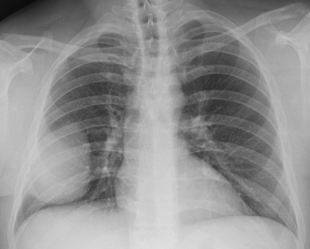
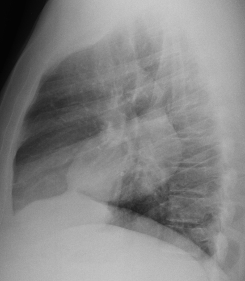
Continuining with interesting cases seen in November, I am presenting the pre-operative chest radiographs of a 44-year-old man scheduled for hand surgery. Would you call the surgeon? What do you think the patient has?
Check the images below, leave your thoughts in the comments section and come back on Friday for the answer.
This pre-op radiograph was shown to me by a technician, who saw the obvious mass in the right lung. A lateral view was taken, showing that the mass is somewhat oval, with a suggestion of a peak in the upper part (B, arrow).
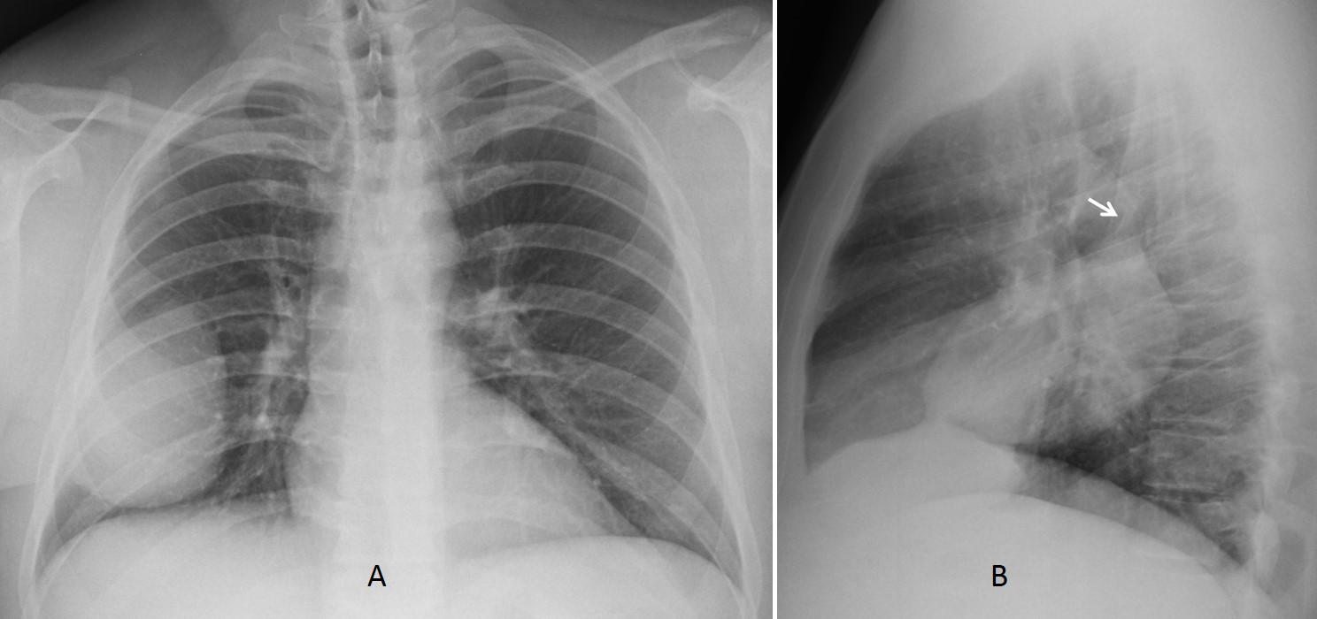
A & B
Unenhanced CT was done at that time, confirming a solid mass (C, arrow), which seemed to be located in the major fissure in the sagittal projection (D, arrow).
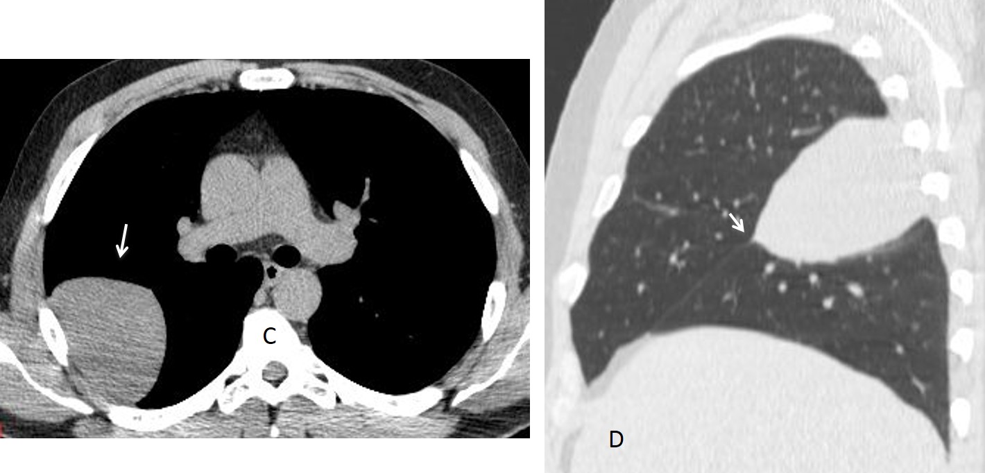
C & D
The obvious diagnosis was an intra-fissural pleural tumour. The surgeon was called and the patient was operated on two weeks later.
Final diagnosis: unsuspected fibrous tumour of pleura in major fissure.
Congratulations to Gus, who was the first to give the diagnosis. Most of you suggested the right location of the lesion, although I disagree with those that mentioned loculated fluid; it is an unlikely possibility in a 48-year-old asymptomatic patient.
Teaching point: I have to emphasise again the importance of the lateral film. In this case it pointed to the right diagnosis, later confirmed with CT.