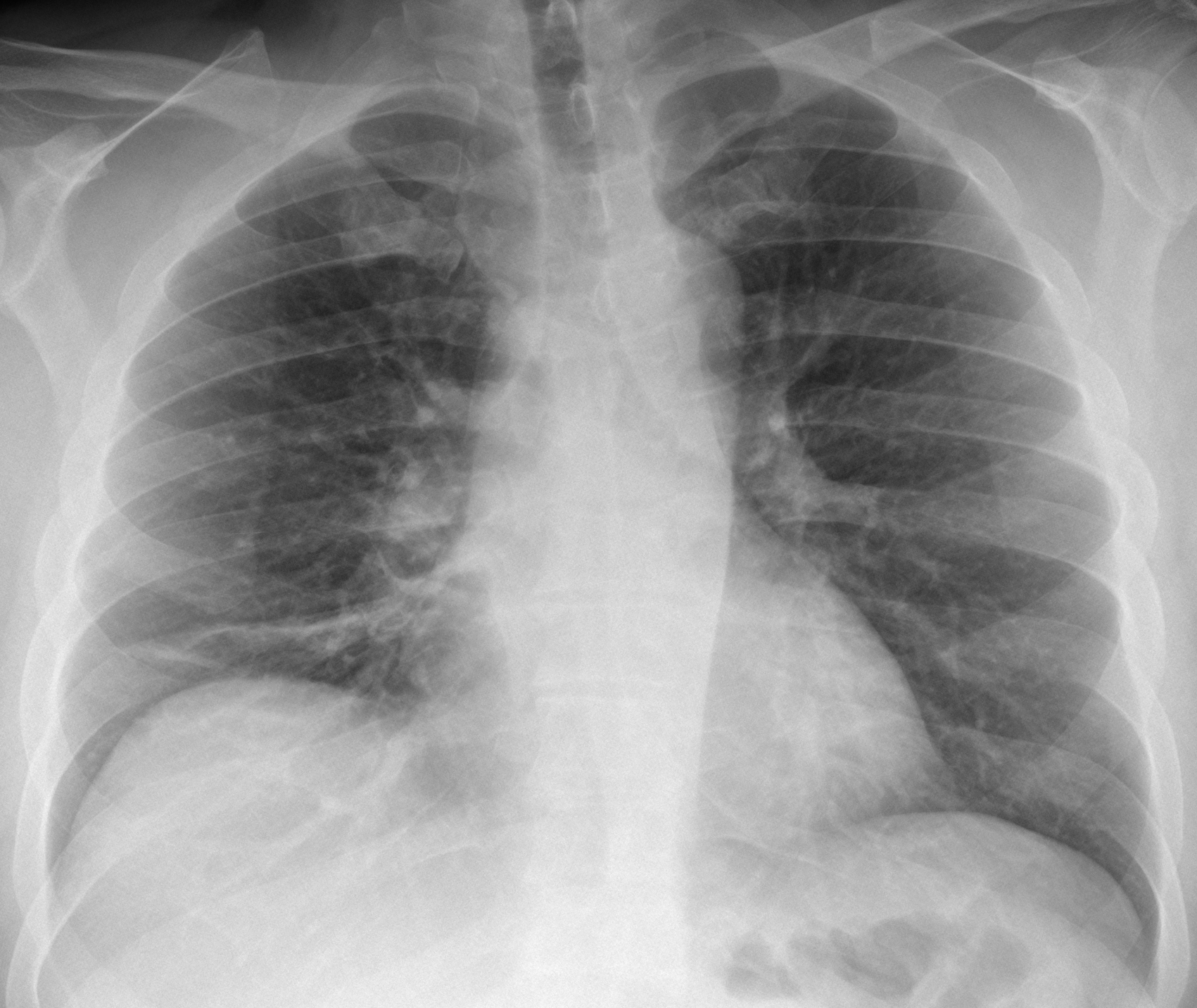
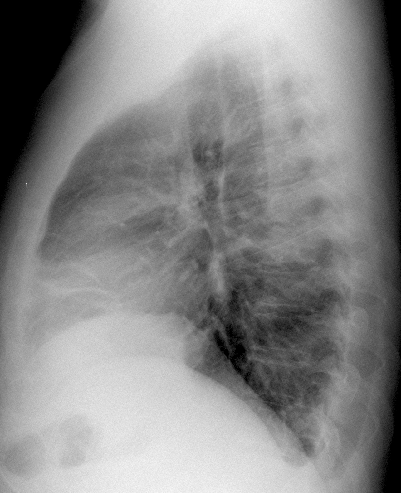
Today we are showing radiographs of a 34-year-old man with malaise and chest pain.
What do you see?
Check the images below, leave us your thoughts in the comments section, and come back on Friday for the answer.
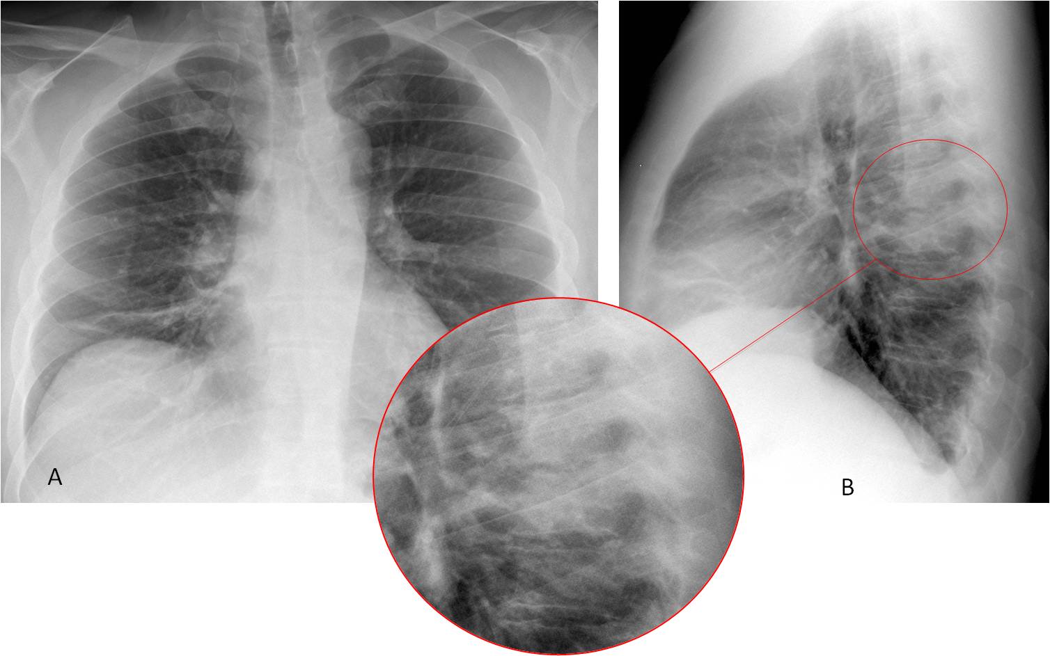
Findings: PA radiograph (A) shows plate-like atelectasis at the right lung base. The right hilum seems increased in density, but it is normal on the lateral projection. The main findings are seen in the lateral view, where the T6-T7 vertebral bodies are more opaque than the ones above or below (B, circle). On close inspection, the intervertebral space is ragged and irregular (insert). These findings are highly suspicious of spinal infection.
Sagittal CT confirms disk involvement (C, circle). Coronal and axial CT demonstrates swelling of the para-spinal soft tissues (D-E, arrows), which are in part responsible for the increased opacity in the lateral chest radiograph. Biopsy demonstrated inflammatory cells and Staphylococcus aureus grew in the culture.
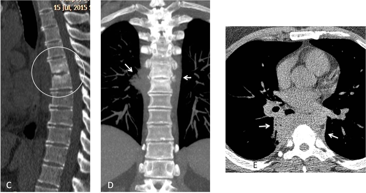
Final diagnosis: Pyogenic spondylodiscitis of T6-T7
Warm congratulations to Murzin, who made an accurate diagnosis.
Teaching points:
1. Remember the importance of looking at the spine in the lateral view
2. Remember that the spine becomes gradually darker as it reaches the lower thorax because there is more lung air superimposed. Seeing a local increased whiteness is suspicious of spinal disease or superimposed lung pathology. It helped to suggest the diagnosis in this case.