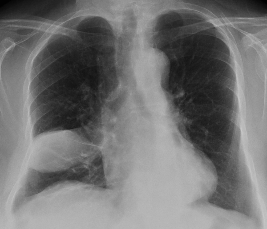
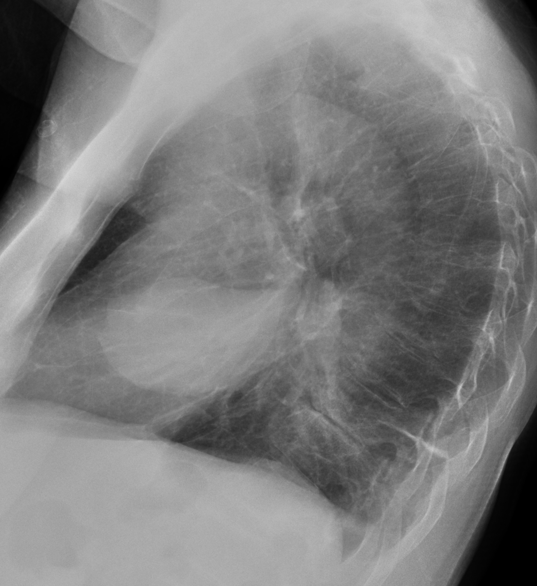
I saw this case in November 13. Radiographs belong to a 72-year-old woman with fever.
1. Infected loculated fluid
2. RML pneumonia
3. Necrotic carcinoma
4. None of the above
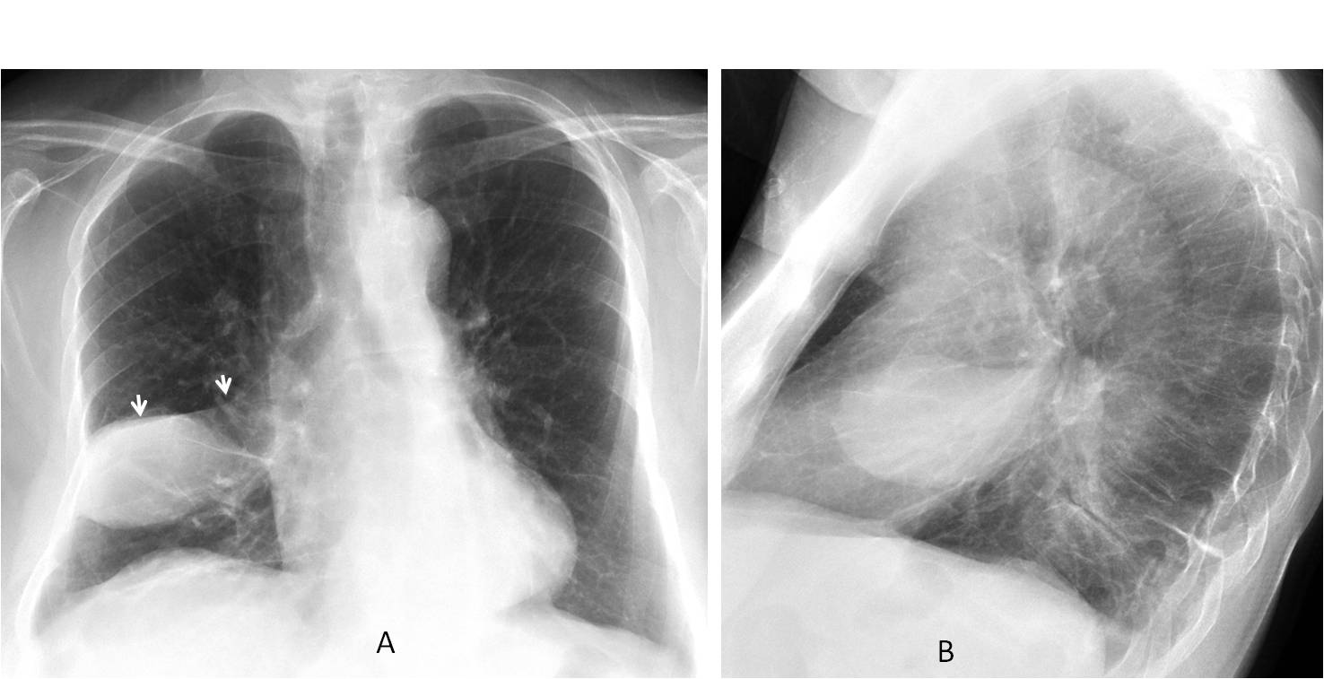
Findings: chest radiographs show a well-defined oval opacity in the right lung, which looks like loculated fluid in the fissure. The clue that disproves this diagnosis lies in identifying the minor fissure, which crosses over the superior portion of the opacity (A, arrows), instead of running through the middle.
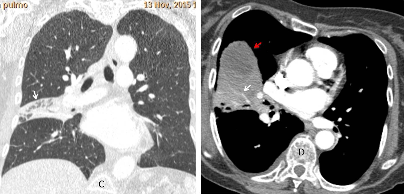
Coronal and axial enhanced CT confirms the presence of lung disease, evidenced by air bronchogram (C, arrows) and vessels within the opacity (D, arrows), with a fluid collection anteriorly (D, red arrow).
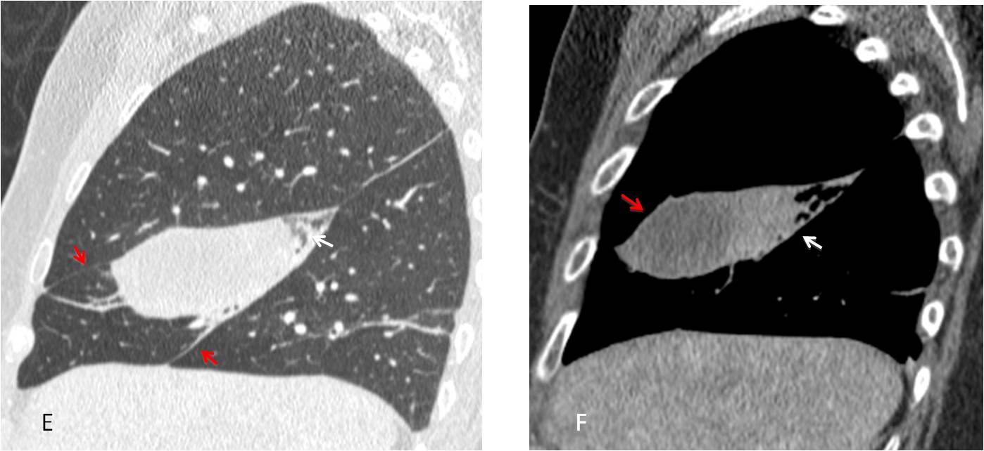
Sagittal reconstruction show the findings more clearly, placing the lung disease in the RML (E-F, arrows), with an abscess anteriorly (F, red arrow). The sharp outlines that simulated loculated fluid are explained by the limiting major and minor fissures (E, red arrows).
Final diagnosis: RML necrotising pneumonia, with abscess
Teaching point: I thought this was a nice Aunt Minnie’s case, in which locating the minor fissure in the plain film leads away from the initial impression of loculated fluid. This is the final case of the year 2015. Back on January 4.
Very happy holidays to all of you!