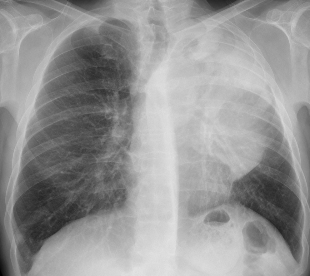
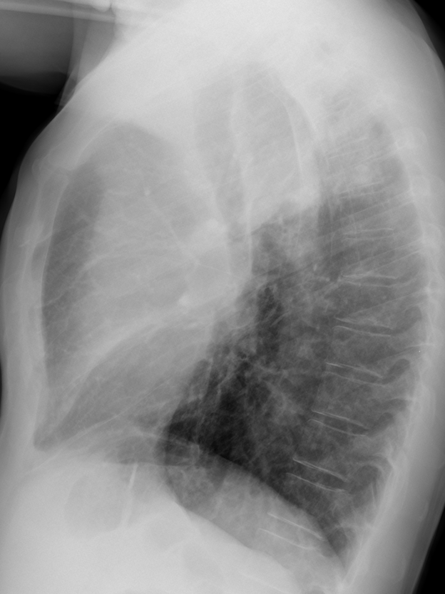
Today we’re showing chest radiographs of a 57-year-old man with chest pain and dyspnoea.
Check the images below, leave your thoughts in the comments, and come back on Friday for the answer.
1. Carcinoma of the lung
2. Thymoma
3. Lymphoma
4. None of the above
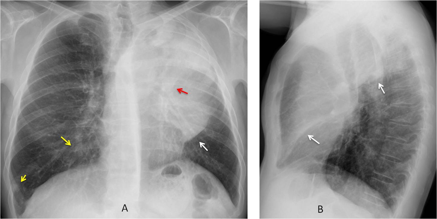
Findings: the PA radiograph shows opacity in the left lung, partially well defined inferiorly (A, arrow). The trachea is displaced and the left hilum is slightly elevated (A, red arrow). This appearance is highly suggestive of LUL collapse with a central mass. Additional findings are several nodules in the left lung (A, yellow arrows), which, in this context, are most probably metastases.
The diagnosis is reinforced by the lateral view, which shows the typical pancake shape of the collapsed LUL, limited by the major fissure (B, arrows).
Enhanced axial and coronal CT confirms the LUL collapse and occlusion of the LUL bronchus (C-D, arrows). Nodular metastases are visible in the right lung (C-D, yellow arrows).
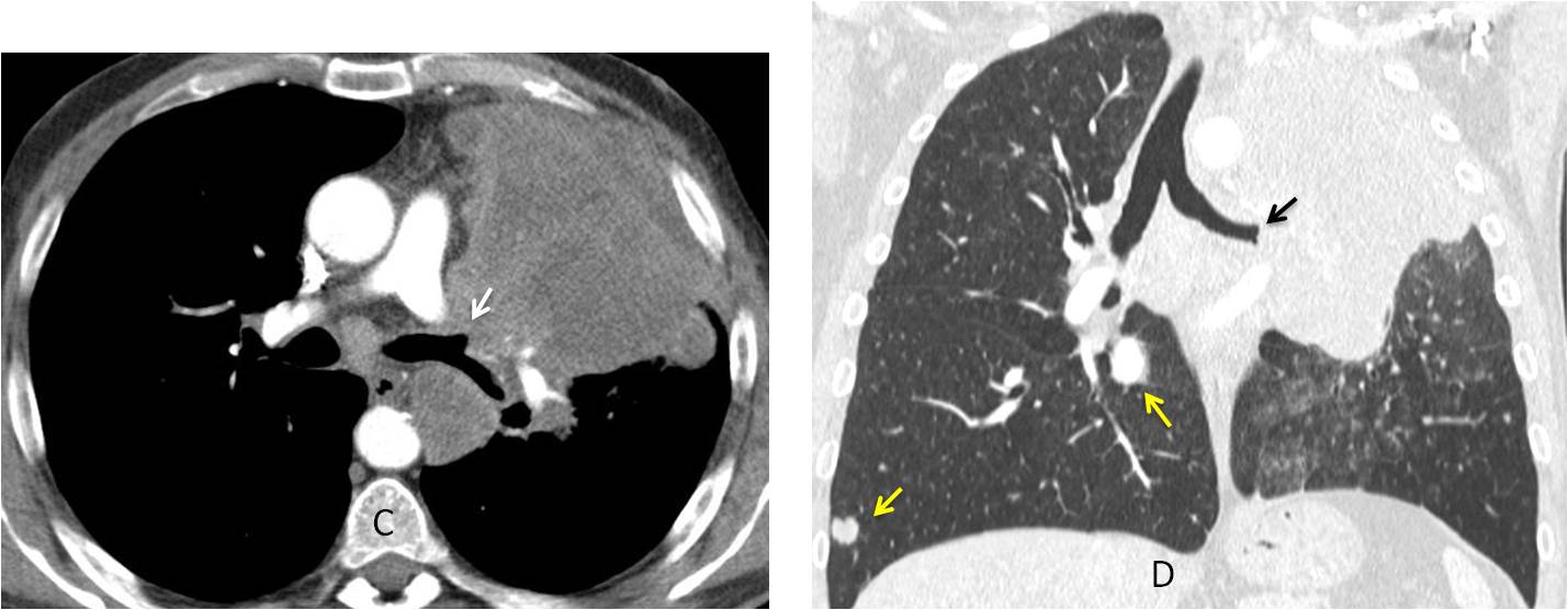
Final diagnosis: carcinoma with LUL collapse and metastases to right lung
Congratulations to MK, who was the first to make the right diagnosis.
Teaching point: diagnosing LUL collapse is reasonably easy for a competent radiologist. Seeing the right lung metastases takes you to the first division. Remember to avoid satisfaction of search!