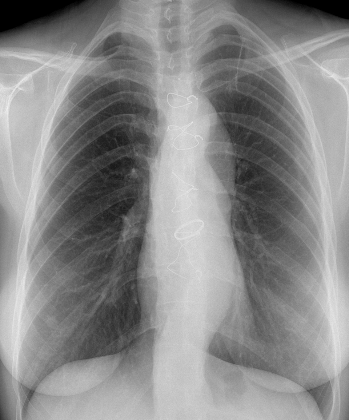
Today we are presenting a routine control radiograph of a 31-year-old woman. Can you guess the reason for the operation?
Check the image below, leave your thoughts in the comments section, and come back on Friday for the answer.
1. Aortic coarctation
2. Aortic dissection
3. Congenital aortic valvular stenosis
4. None of the above
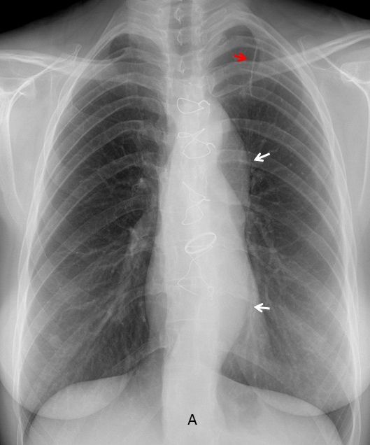
Findings: PA radiograph shows metallic sutures in sternum and a prosthetic aortic valve. In addition the descending aorta is prominent and undulated (A, arrows), which is unusual for the patient’s age. There is an additional finding: surgical staples in the left lung apex (A, red arrow), highly suggestive of previous surgery for pneumothorax.
Of the offered answers, the lack of dilatation of the ascending aorta goes against aortic valve stenosis and aortic coarctation. The combination of pneumothorax, leptosomic chest and undulated aorta suggests aortic dissection in a patient with Marfan disease.
Enhanced axial and sagittal CT (B-C) confirms the extensive dissection. The patient had a spontaneous pneumothorax in 1999, at which time she was diagnosed of Marfan disease. Six years later she was operated on for type A aortic dissection.
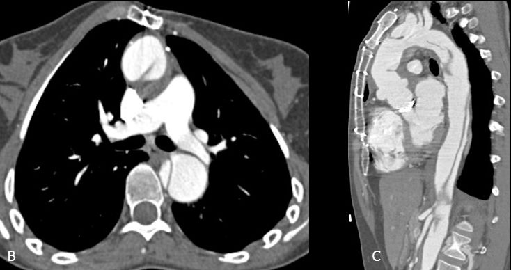
Final diagnosis: Marfan disease with previous pneumothorax and aortic dissection.
Congratulations to the three of you who diagnosed Marfan’s: Mauro, MK and 19Medicus83. Special mention to MK who was the only one that saw the surgical staples and its relationship to pneumothorax.
Teaching point: Although seeing evidence of previous pneumothorax is not needed to suggest Marfan’s, is gives you more surety in your opinion. Avoid satisfaction of search.