Dear friends,
Here’s case 12…
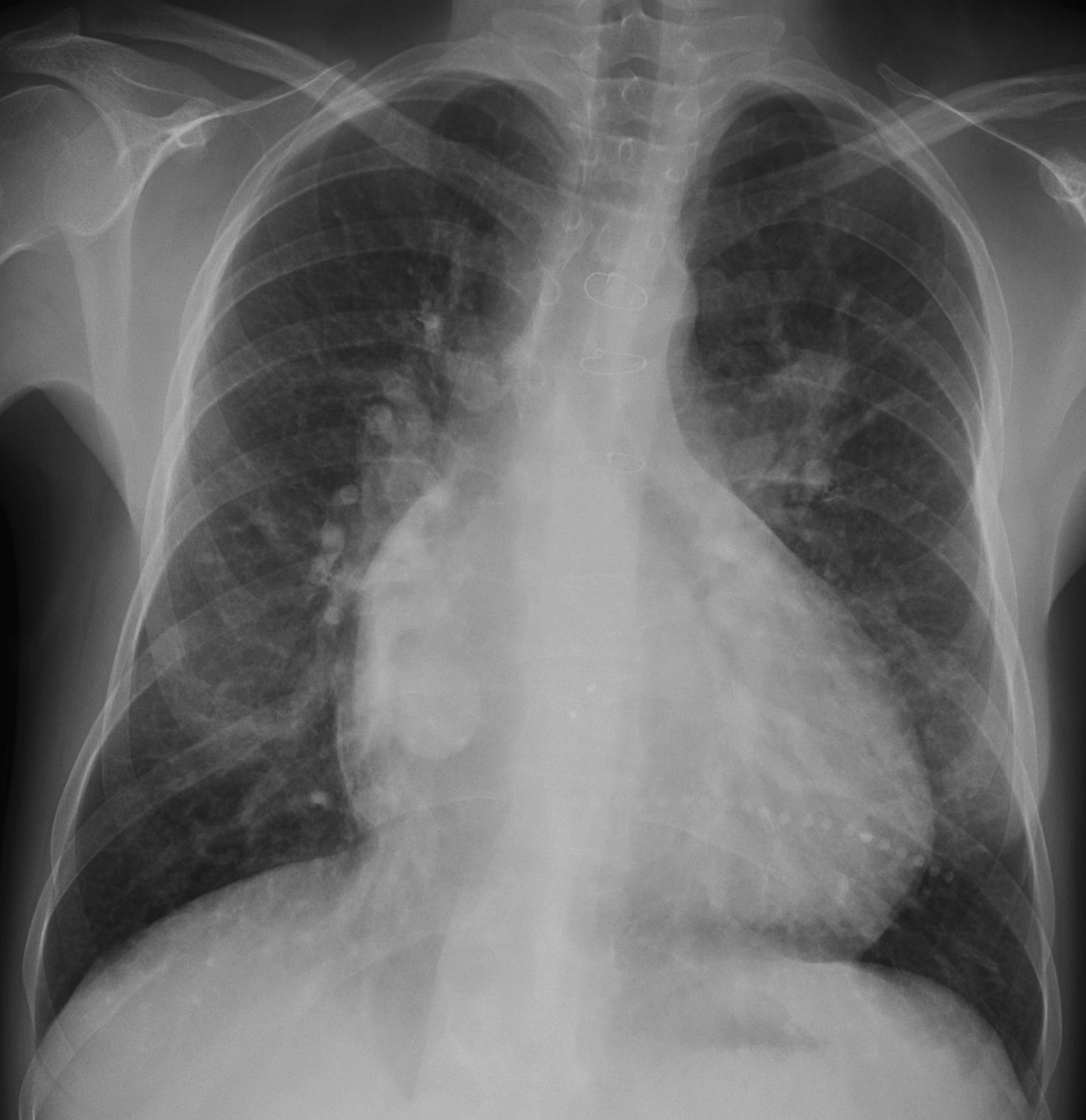
Clinical history: 27-year-old woman, operated on for cyanotic congenital heart disease fourteen years ago.
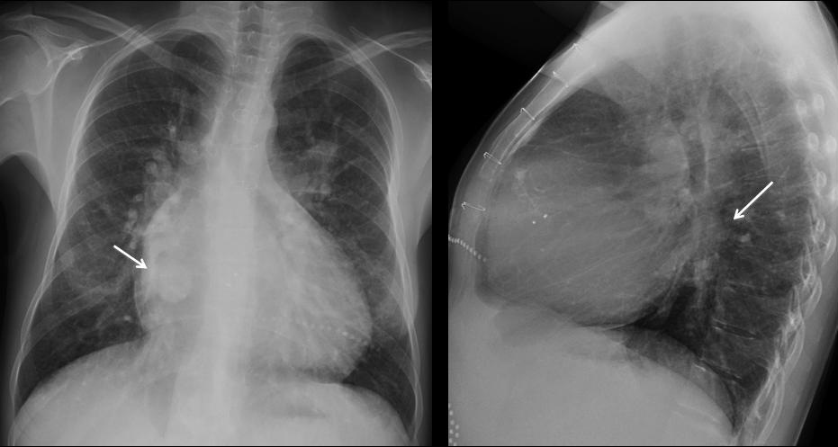
Muppet was intrigued about the rounded opacity in the right lung.
Diagnosis:
1. Hamartoma
2. Hydatid cyst
3. Pulmonary varix
4. Arterio-venous malformation
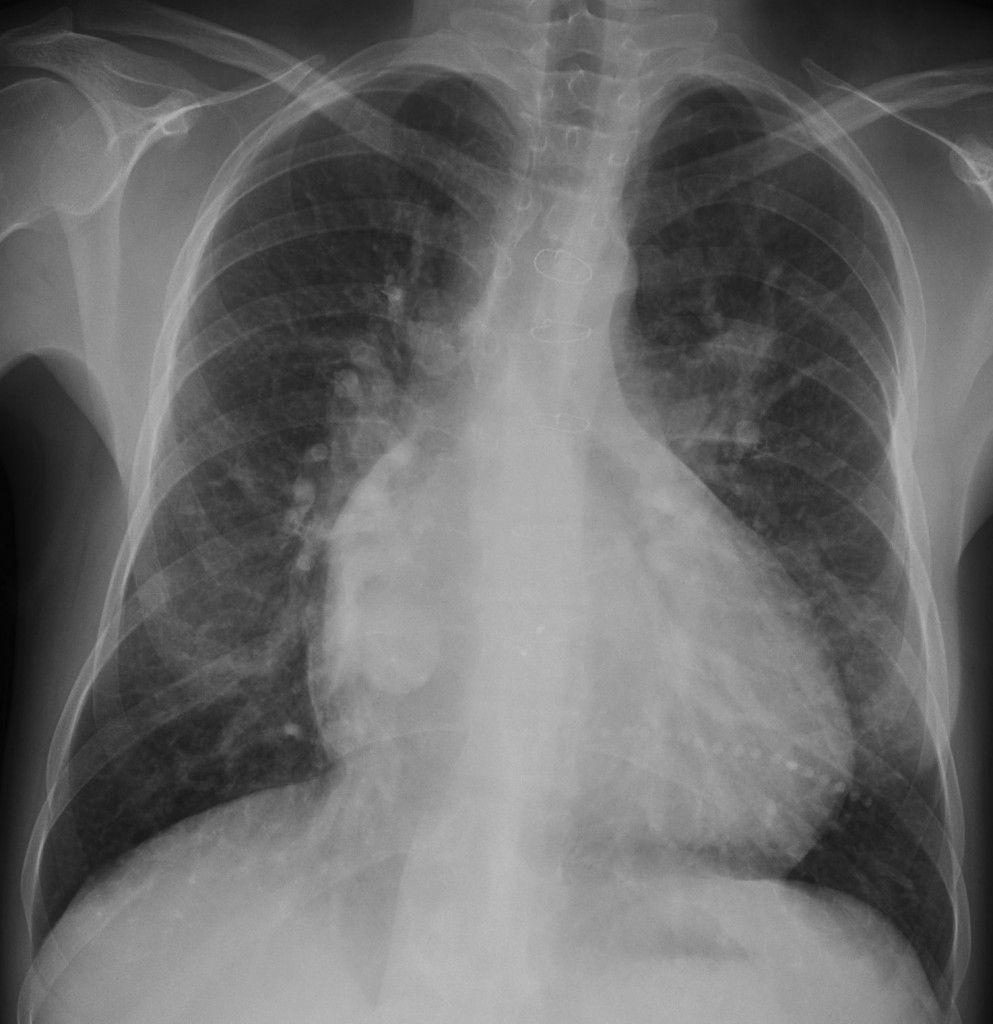
27-year-old woman, PA chest
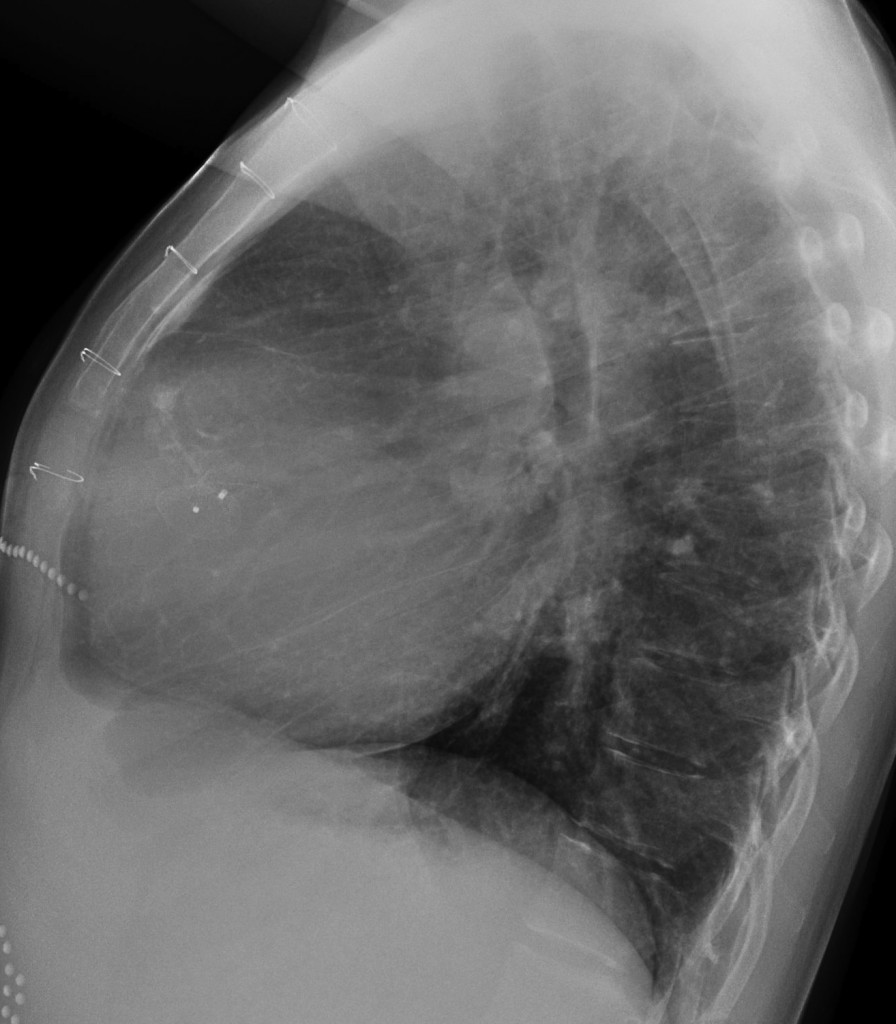
27-year-old woman, lateral chest
Click here for the answer to case #12
Findings: a rounded opacity is seen in the PA view, barely visible behind the heart in the lateral view (arrows). The location strongly suggests an enlargement of the right venous confluent (pulmonary varix). Diagnosis is confirmed with enhanced CT, which shows dilatation of the venous confluent before it enters the left atrium (arrows). A chest radiograph one year earlier showed the same findings.
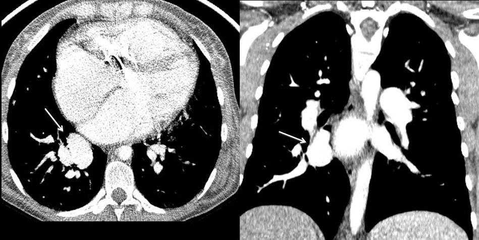
Muppet is an old-timer and has seen a few cases of pulmonary varices in patients with mitral disease, due to increased pressure in the left atrium. Today, rheumatic heart disease is infrequent and varices are rarely seen. In this particular patient the varix is related to congenital heart disease.
Teaching point: it is important to notice the location of a lesion. It may be a determinant in suggesting a likely diagnosis, as in this case.