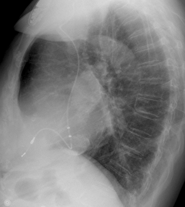
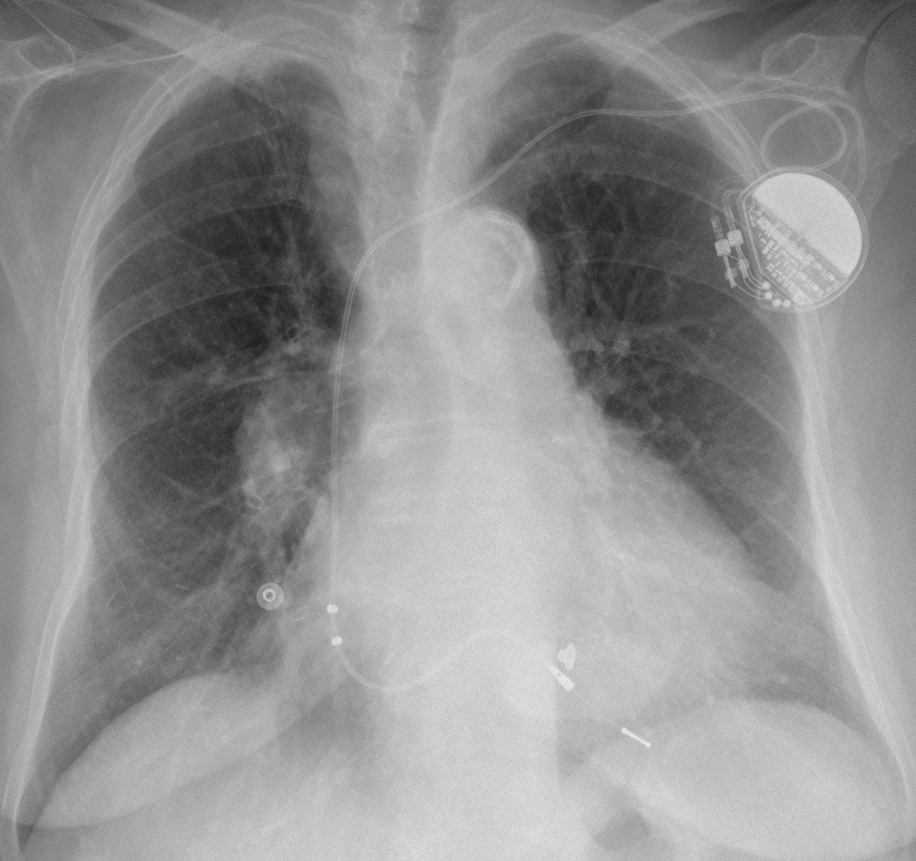
The end of the season is near and I want to show easy cases. Radiographs belong to a 78-year-old woman with dyspnoea. What do you see?
Check the images below, leave your thoughts in the comments section, and come back on Friday for the answer.
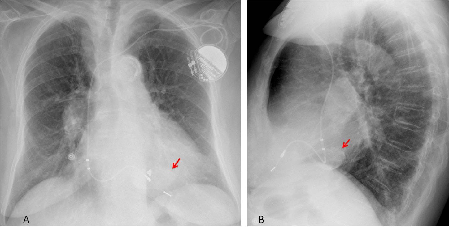
Findings: chest radiographs show an enlarged heart with a pacemaker. The intriguing finding is a rounded cardiac calcification barely seen in the PA view (A, arrow), and more obvious in the lateral projection (B, arrow). The curvilinear appearance may suggest a calcified aneurysm to the non-initiated, whereas in fact it represents a caseous necrosis of the mitral annulus due to liquefaction of the annulus. It is an uncommon variant of the more frequent C-shaped appearance.
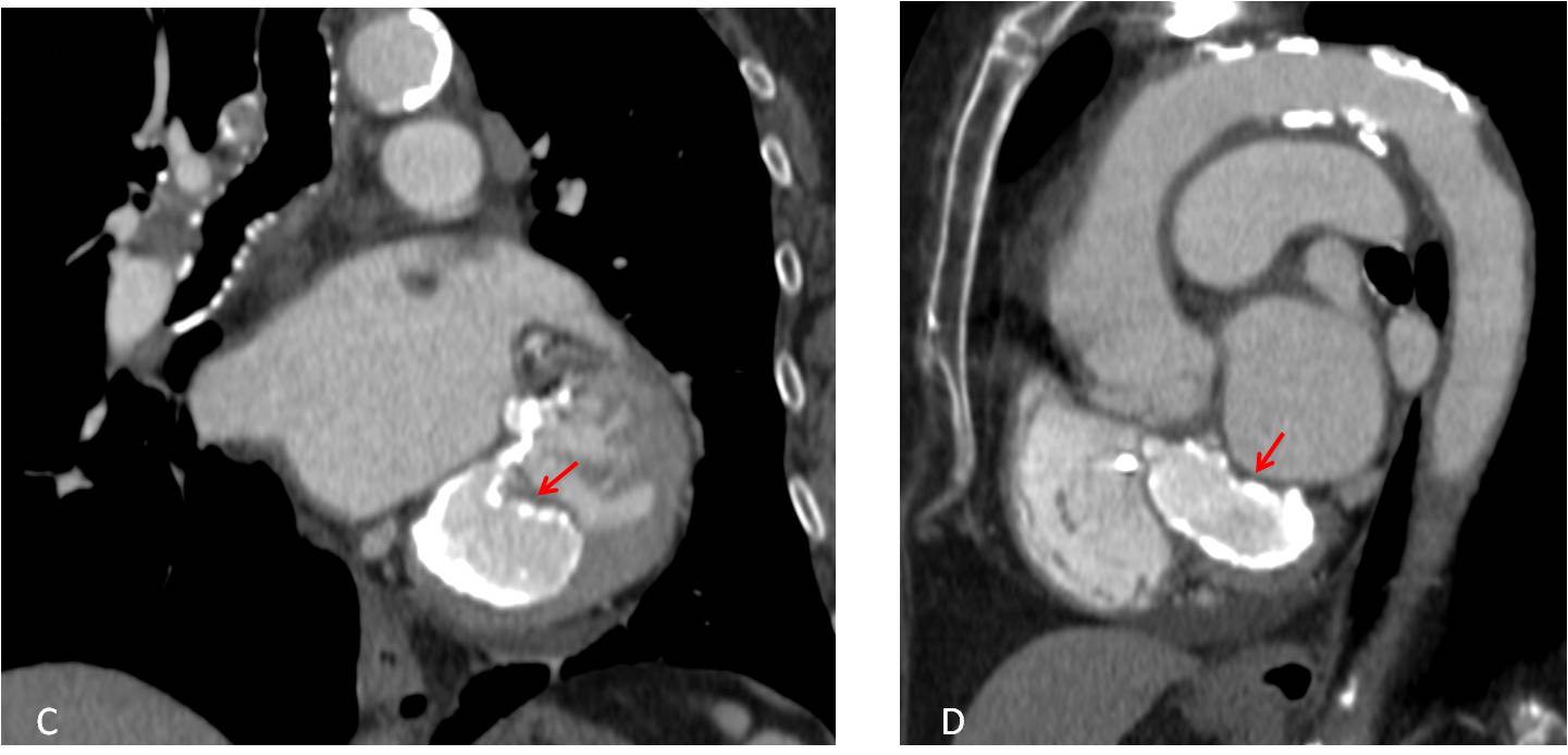
The findings on the plain film are confirmed with the typical appearance in CT (C-D, arrows).
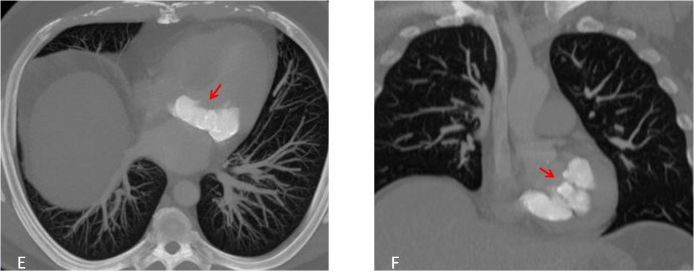
I am showing radiographs of another case seen a few weeks ago, which could be appropriately named as “the mother of all necrosis” (E-F, arrows).
Final diagnosis: caseous necrosis of the mitral annulus
Congratulations to MK, who suggested the diagnosis
Teaching point: know the normal variants. Will prevent you from making erroneous diagnosis and taking unnecessary examinations