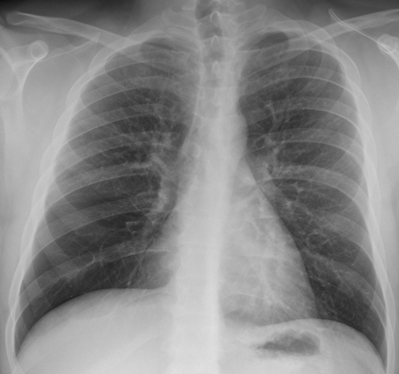
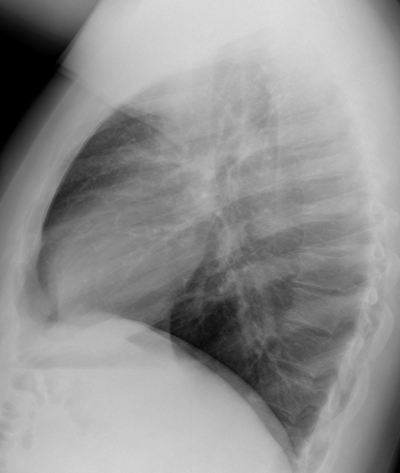
This weeks’ radiographs belong to a 31-year-old male with vague chest complaints. What do you see?
Check the images below, leave your thoughts in the comments section and come back on Friday for the answer.
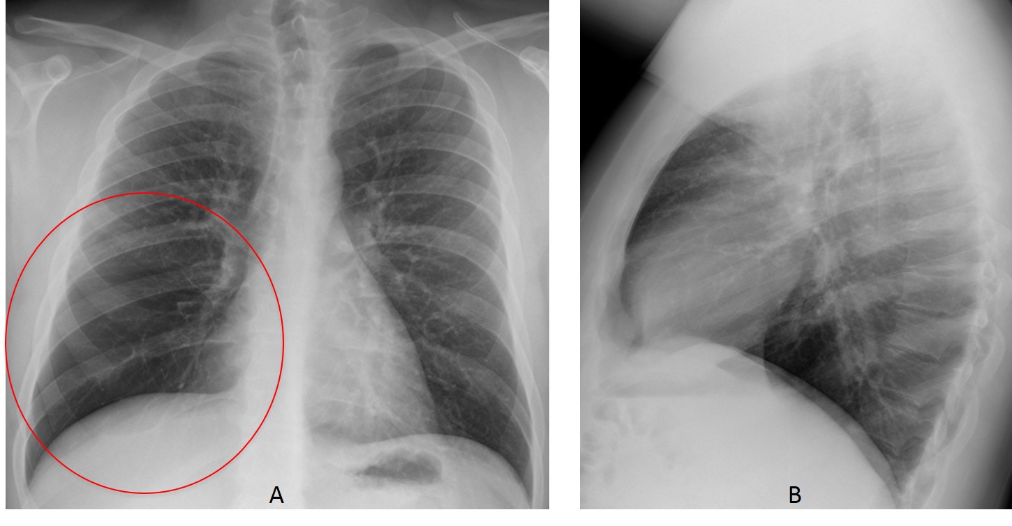
Findings: The lateral view is unremarkable. PA radiograph shows increased lucency of the lower right lung with decreased vasculature (A, circle).
This finding has two main causes: increased lung air or paucity of lung vessels (pulmonary embolism, arterial stenosis). In these cases, the best approach is to take an expiratory film, which will demonstrate whether or not there is air-trapping. If present, it will orient us to an obstructive process, either central or peripheral.
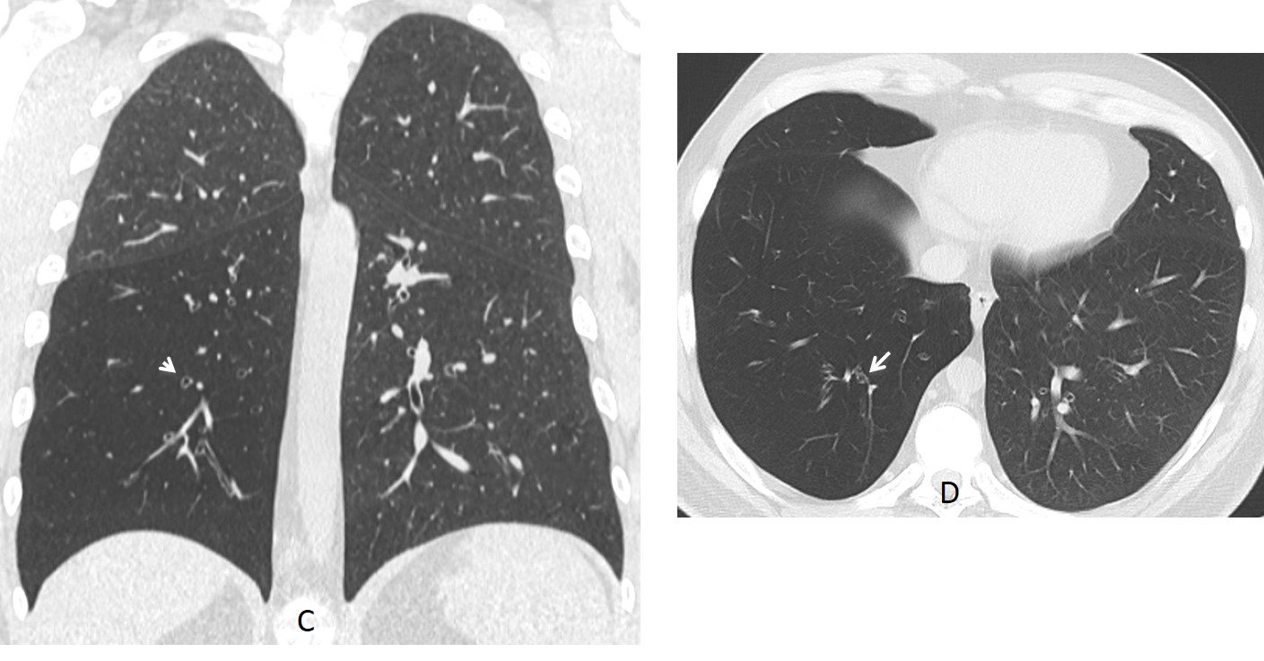
Unenhanced CT confirms the increased lucency and diminished vasculature of RLL and RML. In addition, scattered bronchiectasis are seen (C-D, arrows).
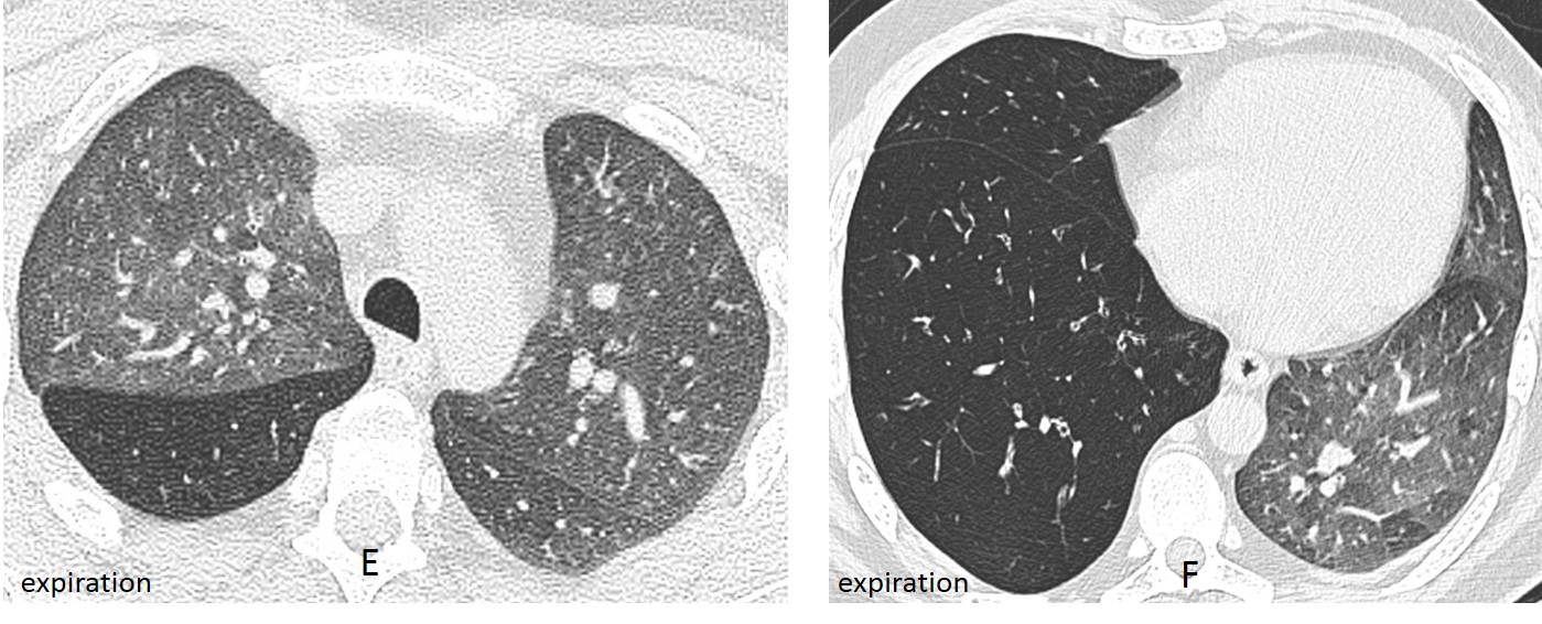
Axial expiratory CTs (E-F) demonstrates air-trapping.
The patient had a history of swallowing a peanut at the age of five years, developing RLL pneumonia at that time. Control radiographs demonstrated increased lucency of the lower right lung over the years.
Final diagnosis: Lobar Swyer-James/McLeod syndrome secondary to aspiration of a peanut in childhood.
Congratulations to TR, who was the first to submit a firm diagnosis.
Teaching point: remember that expiratory films in hyperlucent lung are very useful to orient the differential diagnosis (see Dr. Pepe’s Diploma Casebook case 86)