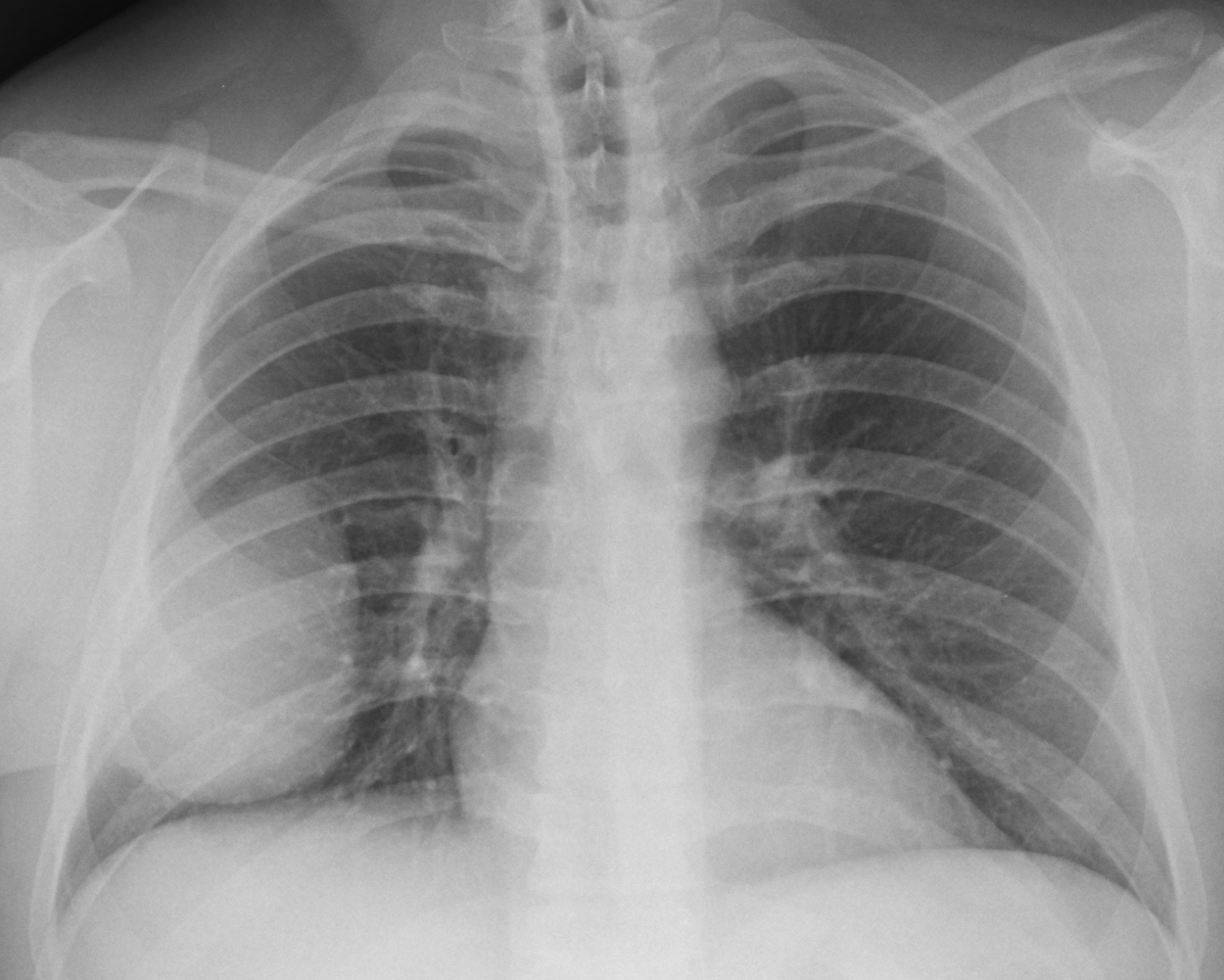
Continuining with interesting cases seen in November, I am presenting the pre-operative chest radiographs of a 44-year-old man scheduled for hand surgery. Would you call the surgeon? What do you think the patient has?
Check the images below, leave your thoughts in the comments section and come back on Friday for the answer.
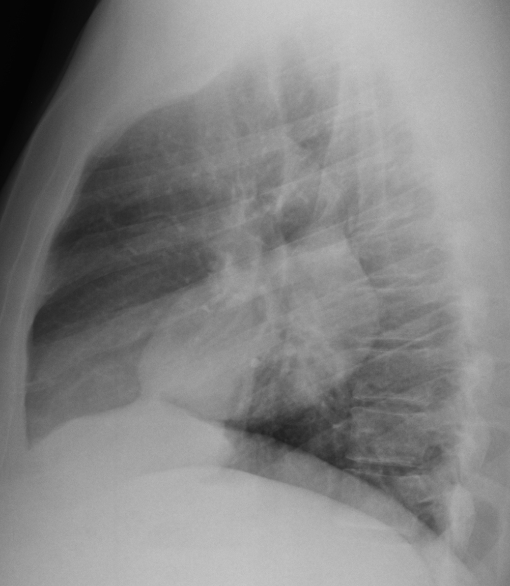
This pre-op radiograph was shown to me by a technician, who saw the obvious mass in the right lung. A lateral view was taken, showing that the mass is somewhat oval, with a suggestion of a peak in the upper part (B, arrow).

A & B
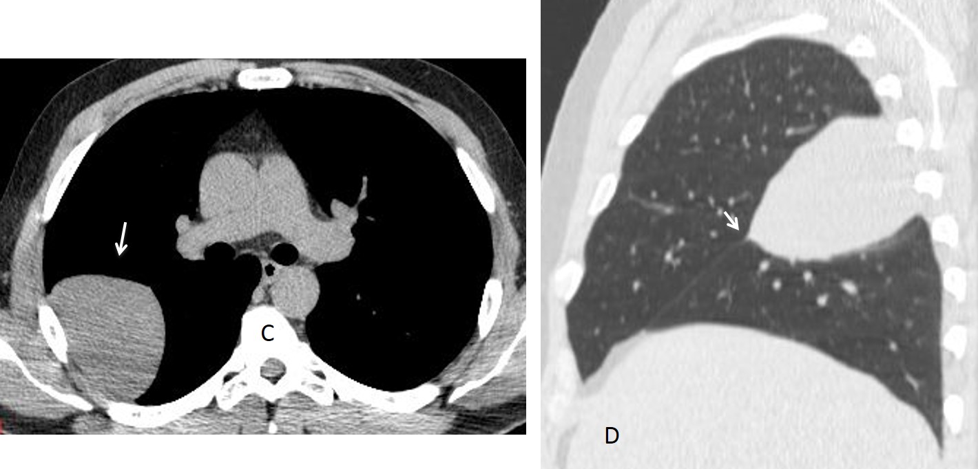
Unenhanced CT was done at that time, confirming a solid mass (C, arrow), which seemed to be located in the major fissure in the sagittal projection (D, arrow).

C & D
The obvious diagnosis was an intra-fissural pleural tumour. The surgeon was called and the patient was operated on two weeks later.
Final diagnosis: unsuspected fibrous tumour of pleura in major fissure.
Congratulations to Gus, who was the first to give the diagnosis. Most of you suggested the right location of the lesion, although I disagree with those that mentioned loculated fluid; it is an unlikely possibility in a 48-year-old asymptomatic patient.
Teaching point: I have to emphasise again the importance of the lateral film. In this case it pointed to the right diagnosis, later confirmed with CT.


pseudotumor or pleural tumor in the right major fissure.
Extrapleural lesion without evidence of aggressiveness.
Good, two answers and it’s only Monday!
loculated pleural effusion in the right oblique scizure.
Maybe encapsulated fluid in the right major fissure.
i think it is loculated pleural efusion…
Why an asymptomatic man would have fluid in the fissure? It usually happens in cardiac failure.
extra chest lesion
empiema]
Chest radiograph PA and Lateral views show a homogenous mass lesion having well defined medial, inferior, anterior and posterior margins. The lateral border of the mass is ill-defined. No rib erosion or calcification or pleural effusion is seen.
I would like to see any previous films if available to see for interval change.
It is a large lesion and the patient is asymptomatic — likely benign pleural mass.
Pleural fibroma (solitary fibrous tumor of the pleura) or pleural lipoma are the possibilities. It can be a (? pedunculated) tumour attached to the lateral pleura or lie in the right oblique fissure.
Is the hand surgery being done for pulmonary osteoarthropathy, ?
since plural fibromas have an association with pulmonary osteoarthropathy.
CT will resolve the issue.
….feliz Navidad,magico professore, da S.Domingo!!!! Sole, mare, rum , sigari ed amore…..ma non ti lascio neppure da Hispaniola ! Lesione pleurica , in asintomatica patologia cardiaca, può’. Essere solo il lipoma od un fibroma nodulare…….Arrivederci all’anno nuovo……..
maybe tumor on scapula?
Tumor fibroso solitario
Mesotelioma
mary christmas!!!
Most probably a benign tumor. If this was malignant, he would have symtomps, probably mets as well.
There’s no effusion, no lymphadenopathy, no mediastinal shifting, no bone destruction etc.
CT must be obtained for better evaulation of the lesion.
Would I call the surgeon? Yes.
Could he still perform the surgery? Probably.
P.S.
Merry Christmas!
🙂
Thnk you. Happy New Year!
encysted pleural effusion in fissure
LFTP or lipoma?
pleural tumor attached to major cisure, probably fibrous estirpe.
No answer on Friday ?
Waiting for the answer.
Sorry, because of the holidays, the answer will be posted next Friday.
Ok. Thanks Sir.
You Spaniards
You got me there! I sleep siesta everyday and dance flamenco at least twice a week
LFTP ? lipoma?
If it was a patient with a known cardiopathy I would say a Pleaural Effusion loculated in the major fissure. Since it is an asymptomatic patient I would say it is more likely to be a benign pleural tumor, probably lipoma.
Happy Holidays
Don’t you think that, in the fissures, fibrous tumor is more common than lipoma?
Extrapulmonary lesion, maybe a pleural solitary fibrous tumour.
Nice case