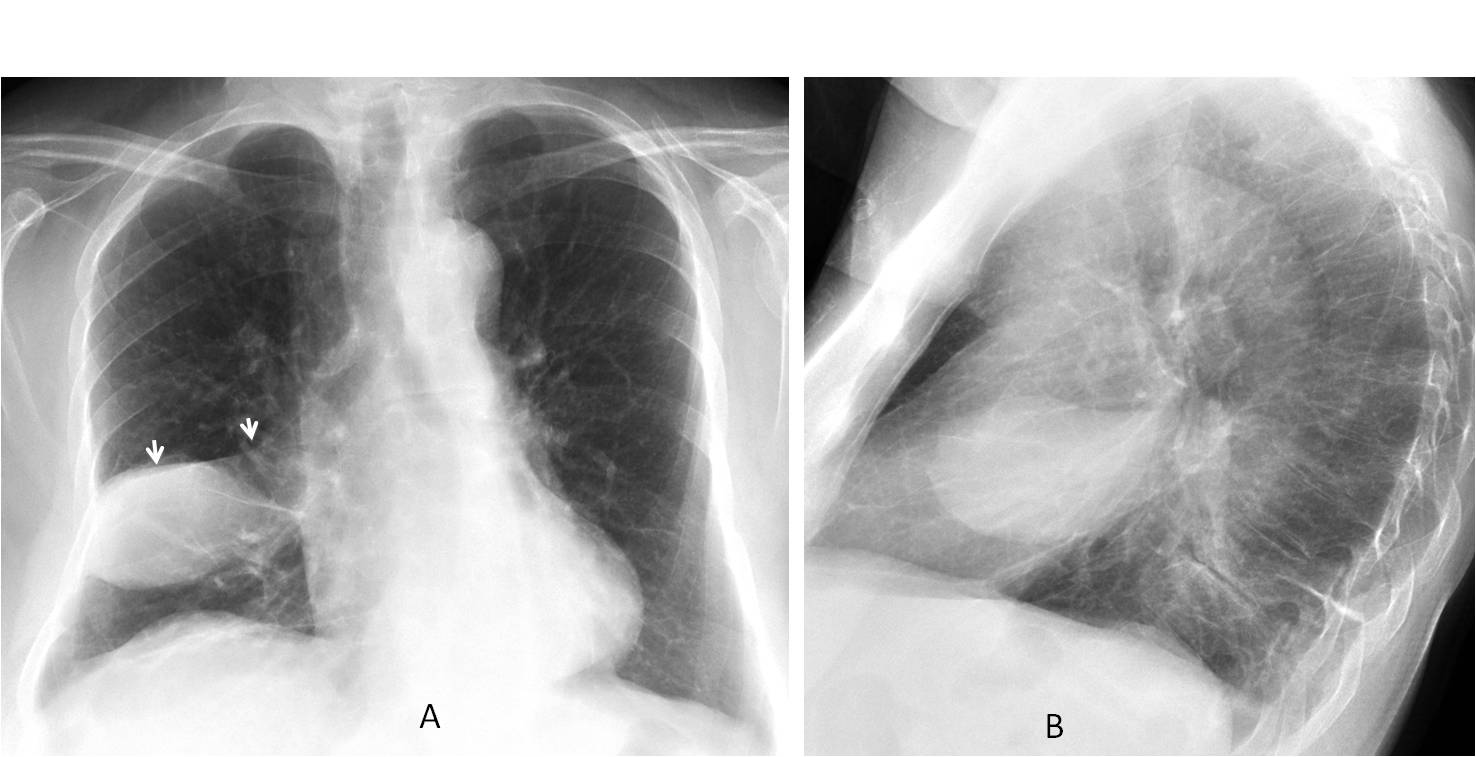
I saw this case in November 13. Radiographs belong to a 72-year-old woman with fever.
1. Infected loculated fluid
2. RML pneumonia
3. Necrotic carcinoma
4. None of the above
Findings: chest radiographs show a well-defined oval opacity in the right lung, which looks like loculated fluid in the fissure. The clue that disproves this diagnosis lies in identifying the minor fissure, which crosses over the superior portion of the opacity (A, arrows), instead of running through the middle.

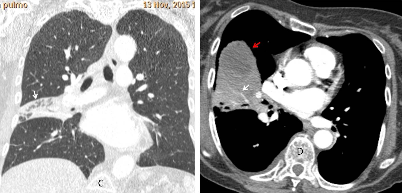
Coronal and axial enhanced CT confirms the presence of lung disease, evidenced by air bronchogram (C, arrows) and vessels within the opacity (D, arrows), with a fluid collection anteriorly (D, red arrow).

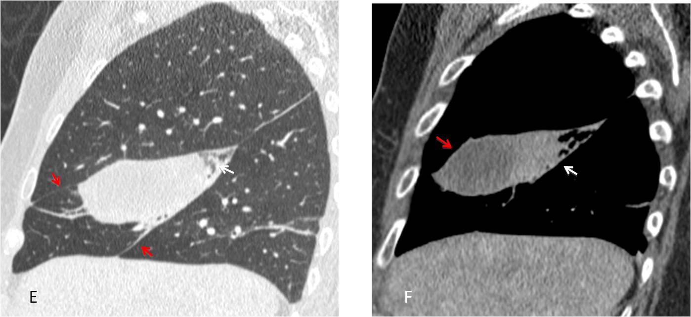
Sagittal reconstruction show the findings more clearly, placing the lung disease in the RML (E-F, arrows), with an abscess anteriorly (F, red arrow). The sharp outlines that simulated loculated fluid are explained by the limiting major and minor fissures (E, red arrows).

Final diagnosis: RML necrotising pneumonia, with abscess
Teaching point: I thought this was a nice Aunt Minnie’s case, in which locating the minor fissure in the plain film leads away from the initial impression of loculated fluid. This is the final case of the year 2015. Back on January 4.
Very happy holidays to all of you!
Infected loculated fluid
1. Infected loculated fluid
Loculated fluid
1. Infected loculated fluid
4.- none of the above.
….mi sembra ci sia un livello idroaereo, nel contesto di una opacità a livello interscissurale e pertanto propendo per una raccolta saccata, infetta….
loculated effusion + segmental collapse consolidation
1. Infected loculated fluid with a collapse of the middle lobe ( loss of volume,…). Besides there is a displacement of the azigoesophageal line maybe because of a hiatal hernia.
1. Loculated fluid
There is a finding in the PA view that goes against loculated fluid. Can you see it?
There is a small amount of fluid in horizontal fisure seen on PA. It is either b or d.
Reasonably close. Answer tomorrow
destruction of the anterior aspect of the 5th right rib?
Ribs are OK
oval soft tissue density superimposed on the minor fissure on both the frontal and lateral views and also there is opacification of the RML abutting the horizontal fissure with elevation of the hemidiaphragm and crowding of the right sided ribs with adjacent abnormal soft tissue components in the chest wall….so goes in favor of malignancy
The appearance is typical of fissural effusion (vanishing tumour)
Necrotic carcinoma?
dear professor, please tell us if a patient has suffered a trauma?
No previous trauma
Infected large bronchogenic cyst. It might communicate with the tracheobronchial tree. Not malignant.
If woman what about breast shadow.. mybe this is out of lung filed.
infected bronchogenic cyst
Non of the above(Hydatid cyst)
….feliz navidad y dichoso ano nuevo,,,MAGISTER !!!!
Grazie il mio amico