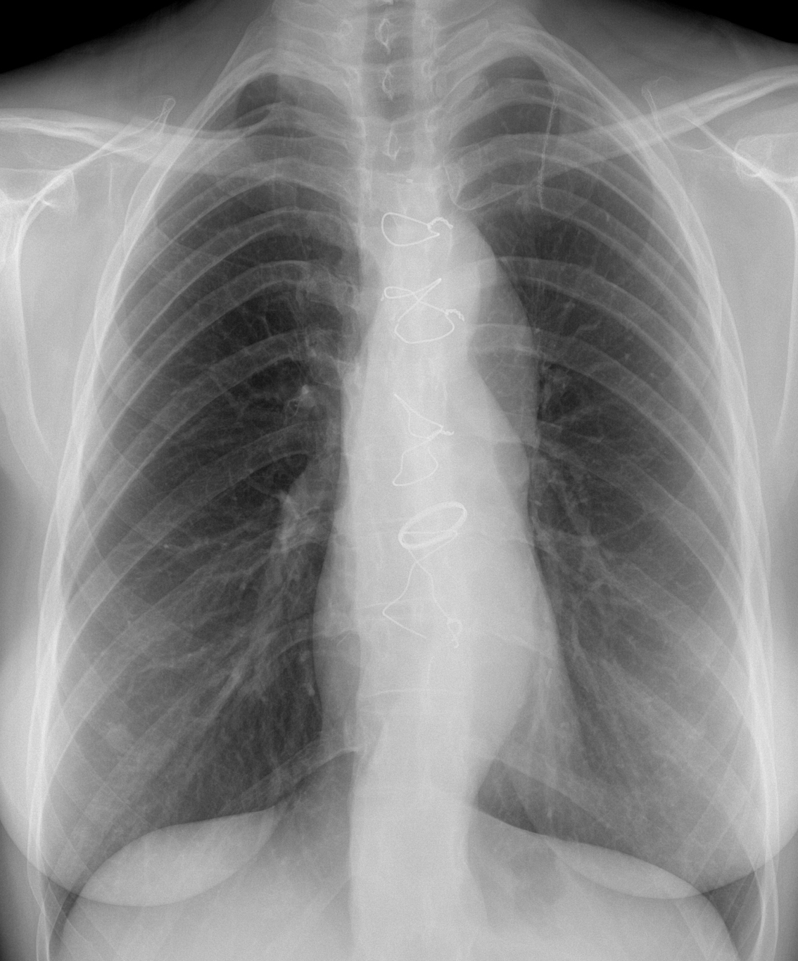
Today we are presenting a routine control radiograph of a 31-year-old woman. Can you guess the reason for the operation?
Check the image below, leave your thoughts in the comments section, and come back on Friday for the answer.
1. Aortic coarctation
2. Aortic dissection
3. Congenital aortic valvular stenosis
4. None of the above
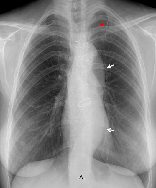
Findings: PA radiograph shows metallic sutures in sternum and a prosthetic aortic valve. In addition the descending aorta is prominent and undulated (A, arrows), which is unusual for the patient’s age. There is an additional finding: surgical staples in the left lung apex (A, red arrow), highly suggestive of previous surgery for pneumothorax.
Of the offered answers, the lack of dilatation of the ascending aorta goes against aortic valve stenosis and aortic coarctation. The combination of pneumothorax, leptosomic chest and undulated aorta suggests aortic dissection in a patient with Marfan disease.

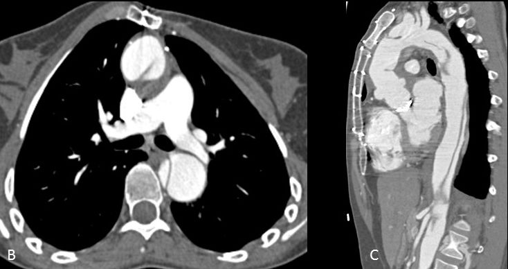
Enhanced axial and sagittal CT (B-C) confirms the extensive dissection. The patient had a spontaneous pneumothorax in 1999, at which time she was diagnosed of Marfan disease. Six years later she was operated on for type A aortic dissection.

Final diagnosis: Marfan disease with previous pneumothorax and aortic dissection.
Congratulations to the three of you who diagnosed Marfan’s: Mauro, MK and 19Medicus83. Special mention to MK who was the only one that saw the surgical staples and its relationship to pneumothorax.
Teaching point: Although seeing evidence of previous pneumothorax is not needed to suggest Marfan’s, is gives you more surety in your opinion. Avoid satisfaction of search.


Hello.
This patient had an aortic valve replacement. I can see that the aorta is enlarged. Putting everything together, I think this patient could have Marfan Syndrome and the valve disease was probably regurgitation.
Metallic aortic a valve.
Elongated descended aorta
Middle sternotomy
Metallic suture in left apical region
Aortic coarctation is not a good idea because of the dilatation of the descended aorta.
For aortic valvular stenosis the ascend aorta is not diltated
Perhaps the apical left suture is because of a bulla surgery (but there are not loss of volume), and the leptosomic thorax appearance would be more factores to think in Marfan syndrome, like Mauro.
….PROF !!!!! Questo non può’ essere il cuore “nativo”…..è’ troppo piccolo…..questo è’ un cuore “trapiantato”….. attento a Dybala…..sara’ il vostro incubo !
It is not common to have a prosthetic valve in a transplanted heart.
If I were you, I would worry about
Messi, Neymar and Suarez.
Hello professor!
My findings are as nearly all are already mentioned:
– middle sternotomy, second and last cerclage are (partly) broken
– metallic aortic valve replacement
– surgical seam in the left apical lung region
– normal, rather small configurated heart
– double contour of the aortic knob and kinking of the descendens aorta
– prominent pulmonary trunk with an outlining contour projected over it
– both lungs and hila look normal
Interpretation seems a little bit difficult for me. Keeping the double contour of the aortic knob in mind I suppose the patient had an typ a dissection affecting the aortiv valve. Consecutively, aortiv valve replacement was needed. Marfan syndrome as the underlying disease seems quite likely.
Non of the above
Hi
I was checking the case again, as it was my first time participating.
Is not usual to find an ao valv replacement ONLY with a type A dissection…
So, i couldnt make 1 dg…
I think answer 1. Aortic coarctation: figure of 3 sign, inferior rib notching. often in association with hypoplasia of the aortic valve (bicuspid aortic valve most common associated defect), probably because it was held prosthetics.