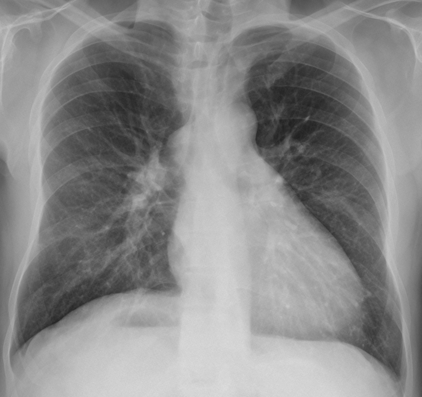
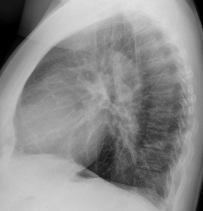
This week I am presenting a case that my wife saw three weeks ago. The radiographs belong to a 45-year-old man, asymptomatic. What will be your diagnosis?
Check the images below, leave your thoughts in the comments section, and come back on Friday for the answer.
1. Enlarged azygos vein
2. Enlarged ymph node
3. Mediastinal mass
4. Any of the above
Findings: there is an oval mediastinal mass at the level of the azygos vein (A, arrow). The pulmonary hila are prominent, and the pulmonary circulation is slightly increased.
The gastric bubble is not visible on the left side because it is located under the right hemidiaphragm (A, red arrow)
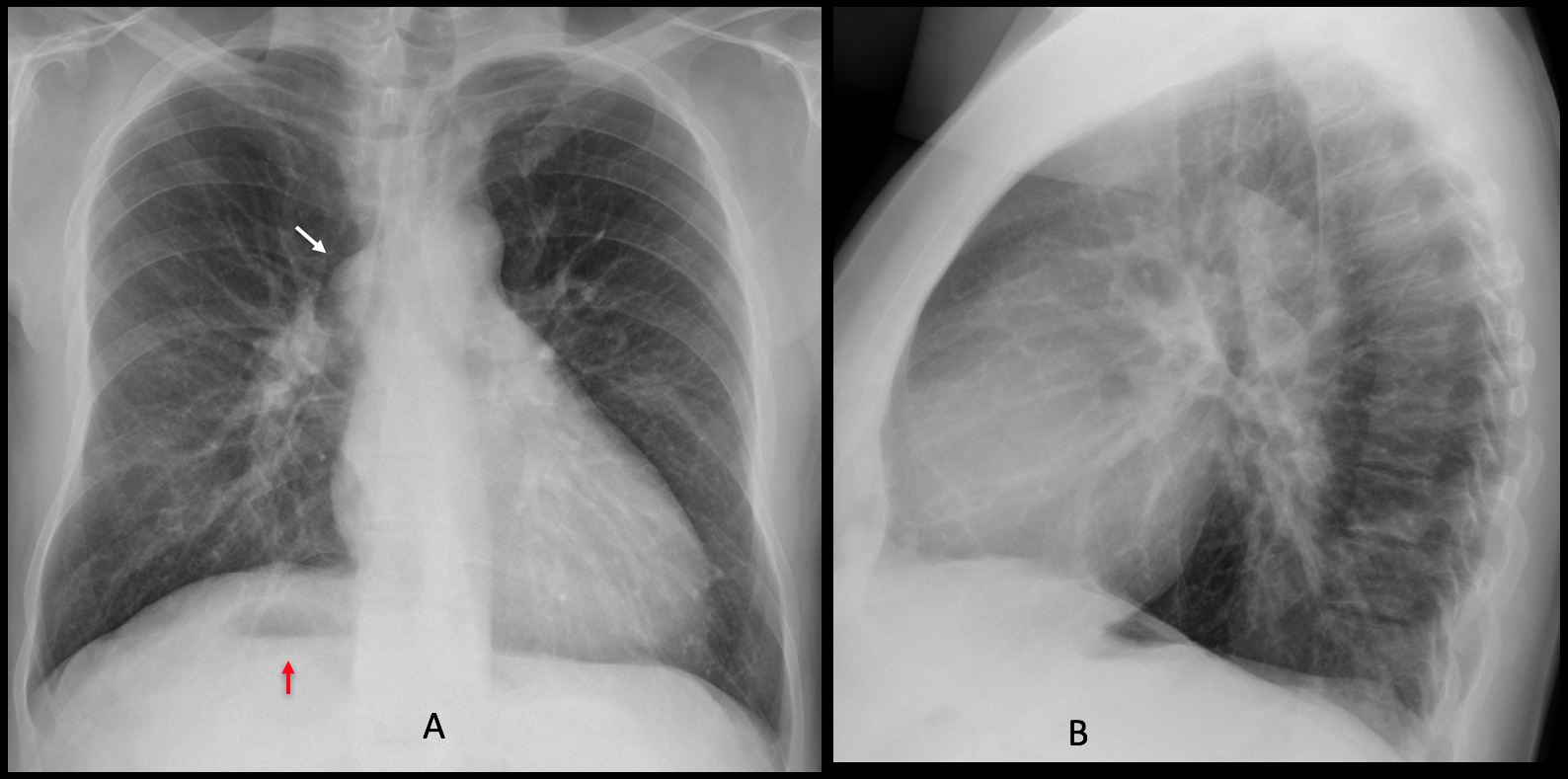
PA chest radiograph six years earlier (C) show identical findings. Radiograph of the abdomen taken at the same time shows the liver in the left upper quadrant (D, arrow) and inversion of the colon.
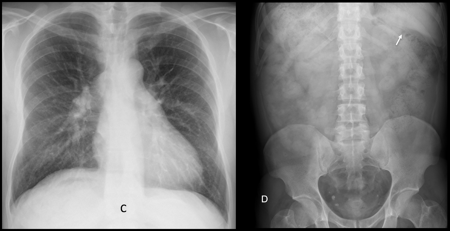
Although CT was not done, the radiographic appearance is suggestive of a normal position of the chest structures and inversion of the abdominal viscera. In this context, the mediastinal mass probably represents an enlarged azygos vein secondary to interruption of the IVC, an abnormality which accompanies the heterotaxy syndromes.
I am no expert in heterotaxy syndrome; reviewing the literature1 I found that this particular malformation is usually accompanied by severe cardiac disease. About 10% of the patients are asymptomatic. This patient is one of the lucky few, although I suspect he has some form of left-to-right shunt.
Final diagnosis: levocardia with abdominal situs inversus
My hearty congratulations to Diogo, who was the first to see the inversion of the gastric bubble, which was the clue to the correct diagnosis.
Teaching point: once again, don’t forget satisfaction of search. Remember to look at the whole radiograph, because the clue to the diagnosis may be away from the main finding.
(1) A case of unusual visceral heterotaxy syndrome Dae Sun Jong and cols (Korean Circ J 2013; 43:705-709)



Good morning!
In the PA and lateral x-ray the azygos vein is enlarged, with an enlarged right hilum respect to the left one.
I am agree with Daniel! Elarged azygos vein because of inferior vein cava agenesia!!
I am not sure about the asymmetry of the hila. They seem the same in the lateral view.
Amigos continuation of ivc?
Azygos** sorry
Enlarge lymph node
Can you differentiate a lymph node from the azygos vein?
Actually it is easy: in the lateral view the lymph node is anterior to the trachea, whereas the azygos is behind it
Double aortic arch?
In my experience, when you see a double aortic arch, the right one is usually higher than the left.
But I may be wrong 😉
I think it could be any of the above choices .
….potrebbe essere un aneurisma dell’aorta ascendente con ipertrofia ventricolare sx.
The azygos vein is enlarged. I can’t see the gastric bubble on the left, but there is some gas on the right. Maybe azygos continuation of IVC as part of an heterotaxy syndrome?
Finally somebody is looking at the whole film!
Congratulations!
Levocardia with situs solitus
Situs inversus sorry…