Dear friends,
Here’s case 12…
Clinical history: 27-year-old woman, operated on for cyanotic congenital heart disease fourteen years ago.
Muppet was intrigued about the rounded opacity in the right lung.
Diagnosis:
1. Hamartoma
2. Hydatid cyst
3. Pulmonary varix
4. Arterio-venous malformation
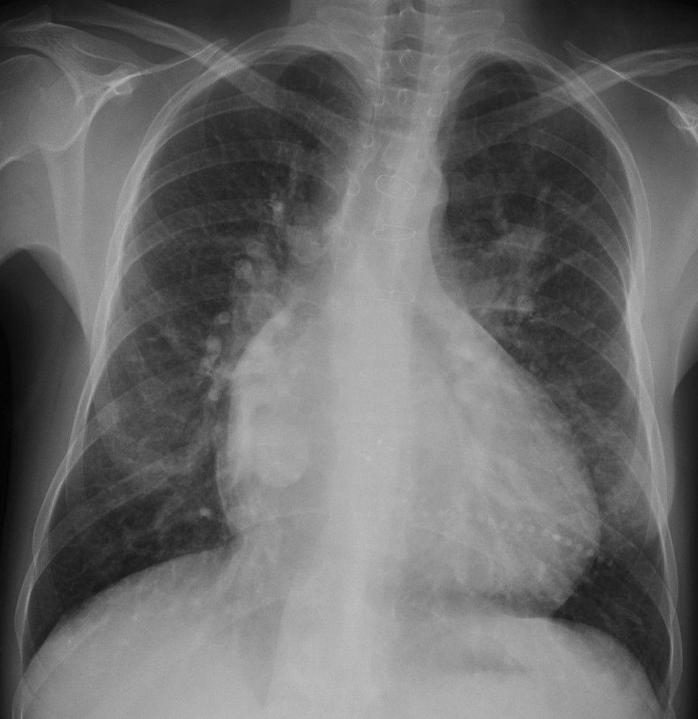
27-year-old woman, PA chest
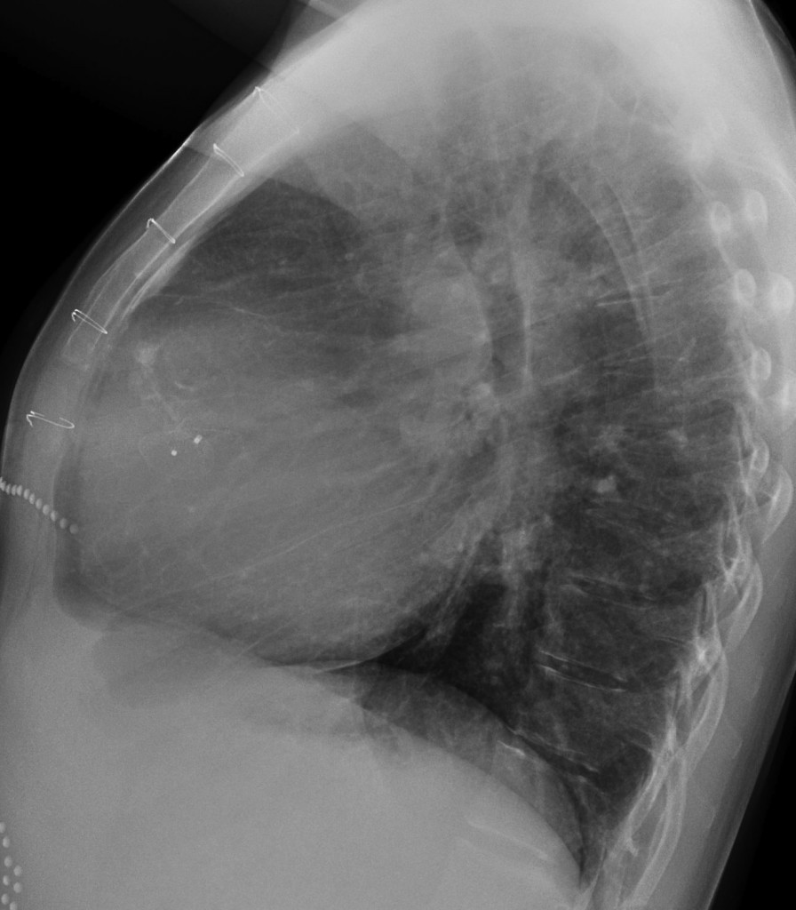
27-year-old woman, lateral chest
Click here for the answer to case #12
Findings: a rounded opacity is seen in the PA view, barely visible behind the heart in the lateral view (arrows). The location strongly suggests an enlargement of the right venous confluent (pulmonary varix). Diagnosis is confirmed with enhanced CT, which shows dilatation of the venous confluent before it enters the left atrium (arrows). A chest radiograph one year earlier showed the same findings.
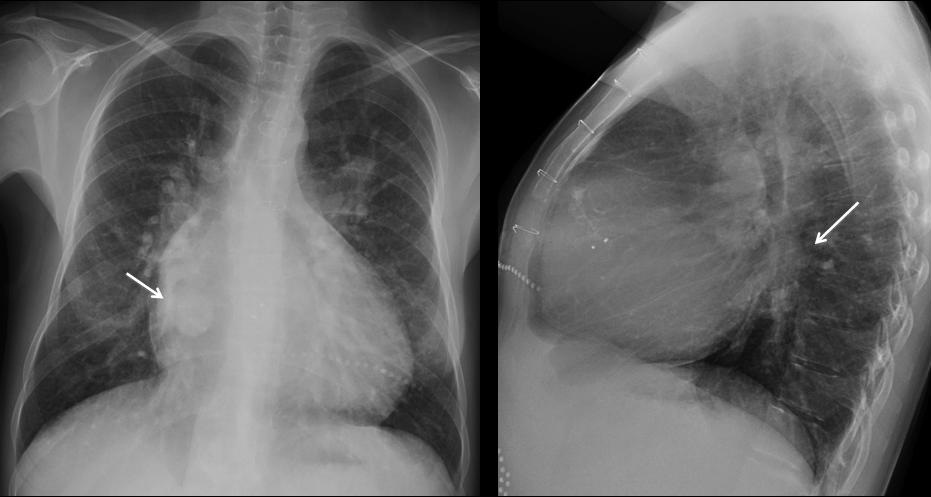
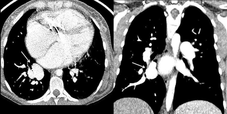
Muppet is an old-timer and has seen a few cases of pulmonary varices in patients with mitral disease, due to increased pressure in the left atrium. Today, rheumatic heart disease is infrequent and varices are rarely seen. In this particular patient the varix is related to congenital heart disease.
Teaching point: it is important to notice the location of a lesion. It may be a determinant in suggesting a likely diagnosis, as in this case.


it’s an Arterio-venous malformation
I think it is hamartoma, as it is smooth well-definde, faintly calcified nodule with its size less than 4 cm. But the only thing that can be against it, is that it is not at the periphery.
3 or 4
Pulmonary varix
-cardiomegaly with prominent pulmonary vascularity and multiple shunt vessels consistent with left to right shunt.should be VSD from the history(relatively early presentation)
-left atrial enlargement and calcification of LA wall.
rounded opacity could be calcified adherent clot.
Plain CT will help
pulmonary varix is the second differential.(because serpiginous vascular structures are seen adjacent to the lesion)
hamartoma
Pulmonary varix
Arterio-venous malformation
Is it really in the right lung? We are not convinced – it is not clearly visible on the lateral unless we are mistaken -???
It could be a lesion of the thoracic wall, we would therefore like to clinically examine the patient.
It is not in the thoracic wall. The Muppet is harsh but fair. And yes, it is not clearly visible on the lateral. I wonder why?
Put your collective minds to work
hamartoma
AVM cauze it is a well defined lesion in contact with some serpiginous opacities coming from the right hilum (vessels) and peripheral smooth calcifications
My second choice its pulmonary varix
Its not hamartoma cause there are not’ popcorn calcifications’ and its a hidric opacity, hamartoma has to contain fat
Hidatic cyst cant be because its the opacity is hidric and has peripheral smooth calcifications ( very very rare in pulmonary hidatic cyst)
Thorough discussion; but has two inexactitudes: not all hamartomas have calcium and not all of them contain fat.And fat cannot be distinguished from hidric opacity in the plain film.
Aside from that, Muppet loves your discussion!
It is not visible on the lateral, how can you be sure it is in the lung?
Could it be a thoracic wall lesion?
pulmonary varix
1.i probably repeat P-A with all clothes off.
cant see that round shadow clear on the lateral,especially cause she had some metal objects on clothes,and sternal sutures too…vote for foreign body on her clothes,buton? 🙂
2.question,why is right paravertebral line so unsharp?
3.but if i take this case seriously,thats in projection of venous ostium in right atrium?
Take the case seriously.No buttons or skin lesions. Muppet does not like to deceive friends. Although he lies occasionally to improve his image!
of course,i think this is a ortoprojection of some vascular structure,some kind of aneurism or varix,its lower then pulmonary artery structure position,so venous origin is possible…ddx.maybe tromb in atrial auricula or posision of some graft?
Varicous dilatation of the right inferior pulmonary vein.
— I think you can see the structure (but with a tubular form) on the lateral view, so it shouldn’t be a nodule: no hydatid, no hamartoma. Too smooth, to round on PA and too tubular on the lateral to be an AVM. It must be a single vascular structure. Its size and its location suggest to me that it is a varicous dilatation of the right inferior pulmonary vein.
Good. Made the Muppet very happy
hamartoma….
AVM
AVM: both A and V pulmonary vessels clearly enlarged, not intrapulmonary nodule -would have a complete border on PA, not only on lower border, can’t see on profile.
i thin it is hamartoma due to two faint calcfic foci seen.
AVM
Una angio-TC è dirimente per D.D. . penso ad una varice polmonare come ipotesi maggiore all’Rx-standard
Muppet dice: Bravo!
AVM
pulmonary varix
I think…
hamartoma
Usually hamartomas show popcorn calcification especially when it is large in side and usually it is peripheral in location which is not the case here.
Hydatid cyst will show evidence of calcification (usually rim calcification) and usually it is in the right mid and lower zone so it could be anther differential especially if the patient travelled to an endemic area and likes carnivores.
Sorry, to my knowledge, hydatid cysts do not calcify when in the lung. They do in solid organs ( liver, kidney, etc.).
Other than that, your discussion is good. Wait for the answer which will be coming soon.
regarding the history , round en face lesion on PA – tubular on letral film – parietal calcifications – location – the aspect of all lung and heat shadow (some signs of pulmonary venous hypertension ) all these made the diagnosis between pulmonary varix (most propabil) and AVM
it was heart shadow sorry..
Nobody is perfect!
Confluence of pulmonary veins