Dear friends,
Muppet was grumpy and decided to spent the Easter vacation in the Costa Brava (I suspect he had a secret date with Miss Piggy). He sent the above picture and the following case:
Seventy-five year-old woman, with pain in the back
Diagnosis:
1. Metastases
2. Lymphoma
3. Paget’s disease
4. None of the above

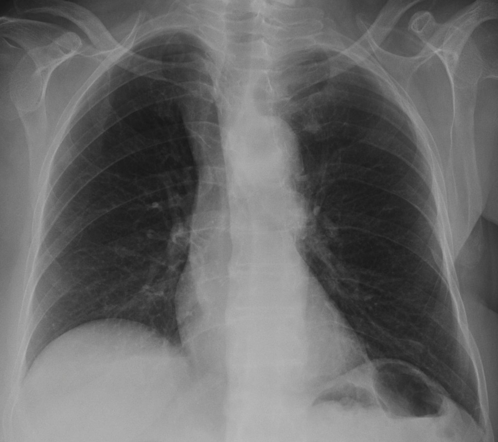
Seventy-five year-old woman, PA chest

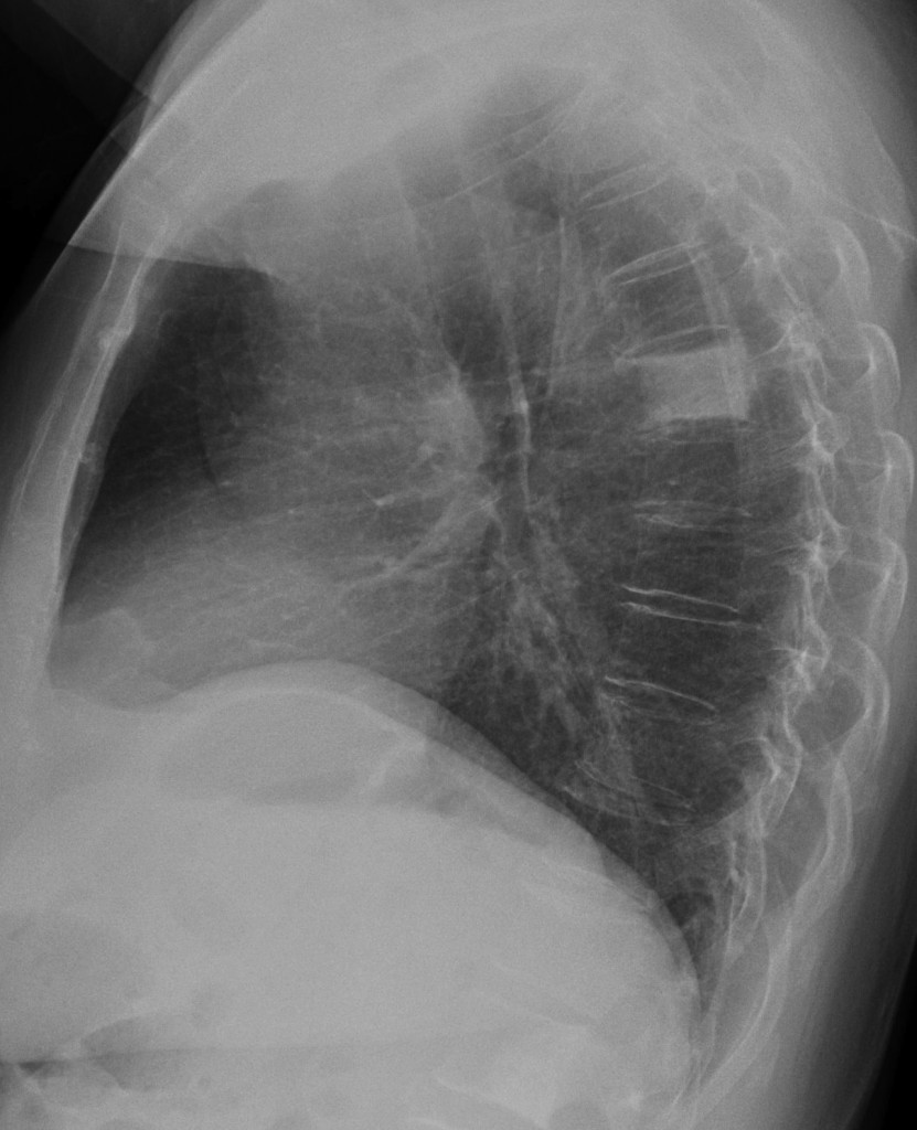
Seventy-five year-old woman, lateral chest
Click here for the answer to case #17
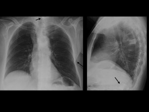
The lateral chest shows an obvious ivory vertebra. The main differential diagnoses of ivory vertebra are metastases, Paget’s disease and lymphoma, and one of you even offered a reference (Muppet applauds Marius). Looking carefully, there is a second osteoblastic deposit in one of the lumbar vertebrae (arrow) and a third in the cervical area in the PA chest film (arrow).

These findings suggest metastases. Once the diagnosis of osteoblastic metastasis is considered, think of two organs: prostate in the male and breast in female. Looking at the breast shadows; the left one is missing. The telltale signs are the triangular shadow of the mastectomy in the soft tissues of the left hemithorax (arrow) and the decreased opacity of the left lower lung. Muppet is very surprised that none of you mentioned the absent breast.
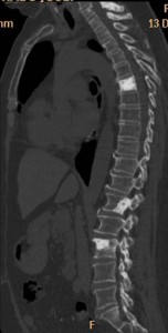
Final diagnosis: osteoblastic metastases from operated breast carcinoma. Sagittal CT reconstruction confirms the findings.

Teaching point: always consider a rare manifestation of a common disease rather than a common manifestation of a rare disease. It works most of the time. In this particular case it would have lead you to search for the missing breast.


paget diseases
Ivory vertebra at T7. To sum up http://radiology.rsna.org/content/235/2/614.full.pdf : Met, Paget, Lymphoma > anything else. For Paget- there is no expansion / there would rather be the picture frame. I don’t see any evidence of hillar or right paratracheal ADP.
Osteoblastic lesion occupying the postero-inferior part of L2 body.
There also seems to be a expansile mixed lesion at the inferior angle of the left scapula.
So i’m leaning towards metastases. Hope it’s not some weird carcinoid 🙂 I’m waiting for answer. That’s all i got right now.
Trachea is pushed to the right by a dilated aorta.
lA SCLEROSI OSSEA è OMOGENEA E NON SI ACCOMPAGNA A DEFORMAZIONE E-O COLLASSO SOMATICO: PERTANTO è DA RIFERIRE AI PAGET. nON PUà PERTANTO ESSERE Nè UN LINFOMA NE UNA METASTASI. nON MI RISULTRA CHE CI SIANO ALTRE CAUSE DI VERTEBRA ” D’AVORIO”. è STATA DOSATO LA IDROSSIPOLINA URINARIA??
urinary hydroxyproline was not evaluated
Also saw the permeative pattern on the left scapular inferior angle and I’ll go for mets.
i think it is metastasis, the findings of pagets disease is not seen as well as enlarged hilar LNs that suggests lymphoma>
Cadaques 😉 the top picture. As far as the other two are concerned I would vote for four. Greetings to Mr. Muppet. Hope he visited Mr. Dali’s house 😉
Well, out of two, you got one answer right. Muppet did not visit Dali’s house. He is a fan of Goya.
One more try then, mets:
-breast cancer
-GI tumour, carcinoid
-bladder carcinoma
or lymphoma (less probable) – lack of lymphadenopathy
Did you look at the breasts?
This is an Ivory vertebra. The three main differential diagnosis for an Ivory vertebra are: blastic metastasis, Paget disease and Hodgkin’s lymphoma. The normal concavity of the vertebral body is unaltered and there is no bone expansion or squaring off of the vertebral body. Also, there is no hilar or mediastinic adenopathy. So I think the most probable diagnosis is metastases. There is also an enlarged thoracic aorta.
I’ ll go for “osteoblastic metastases” too. Considering patient’s age, the absence of lymph nodes and “picture frame” sign, it seems like the best choice 🙂
Osteoblastic lesions occupying the entire body of TH6 (ivory vertebra), part of the body of L1 and the inferior angle of the left scapula.
Small osteoblastic lesions are seen in the sternum, in the 3rd right rib and (maybe) in the 1rst left rib.
I vote for 1. Metastases
In L1 the osteoblastic lesion is occupying also the right pedicle.
more likely Lymphoma
1. Osteoblastic Metastases, unless this patient had a previous osteolitic Mets treated with QMT/RDT and now it seems like osteoblastic.
In the other hand there is also a nodular image projected in the left upper lobe, that I am not able to identify in the lateral view, and it seems like there is an asimmetry of the left hilium ( more dense and prominent??adenopathy?. No donut sign).
Hi,
Trachea shifted to right, raised right hemidiaphragm (blunting of anterior cp angle on right side – lateral view)- ??right lowerlobe collapse, a circular transluscent shadow in the lateral view- is it a mediastinal mass?, Rt. apical shadowing and lastly the homogenous radiop opacity of the lumbar vertebra. Am trying to correlate the findings…malignancy?cyst?mediastinal adenopathy? Subpulmonic effusion…Arrow in the dark…
You are making it too difficult. The main finding is the ivory vertebra. What causes it? Are there any other findings that may help you?
After the solution, things appear to be more clear!
It most likely to be a blastic metastasis
http://virilityexreviews.jimdo.com/ Thanks for that awesome posting. It saved MUCH time 🙂
Sir could you give some light on the dilated aorta ? The trachea is pushed to right. Waiting eagerly for your comment.