This case was provided by my good friend Alberto Villanueva, who is a fan of Real Madrid. Aside from that, he is normal.
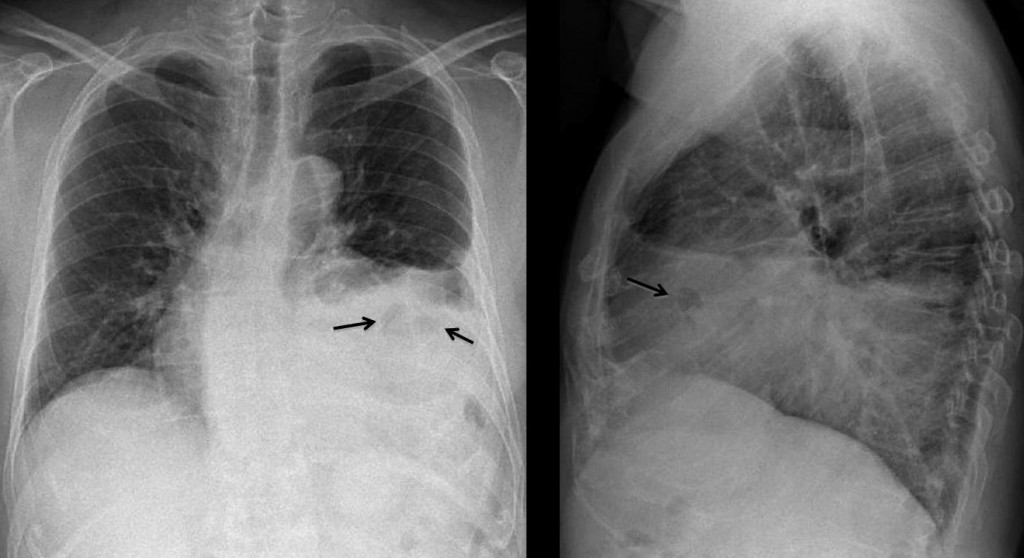
Preoperative chest in a 68-year-old man with a 6cm abdominal aortic aneurysm.
1. Leaking aneurysm
2. Fibrous tumor of pleura
3. Chronic empyema
4. None of the above
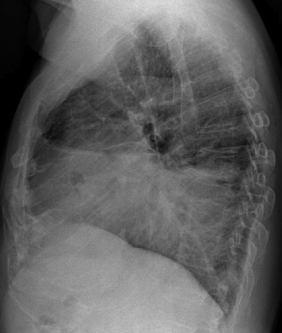
PA and lateral chest show a large opacity in the lower lung field, with an irregular upper border. There is blunting of the costophrenic angle and areas of air-density within the opacity (arrows), which resemble bowel loops.

Once bowel loops are suspected, the choices are between hernia and diaphragmatic paralysis. An elevated diaphragm usually has a concave, well-defined border, whereas hernias have an irregular and flatter contour. If previous trauma is elicited, traumatic hernia should be ruled out. This patient had a severe accident fifteen years earlier and had been asymptomatic since.
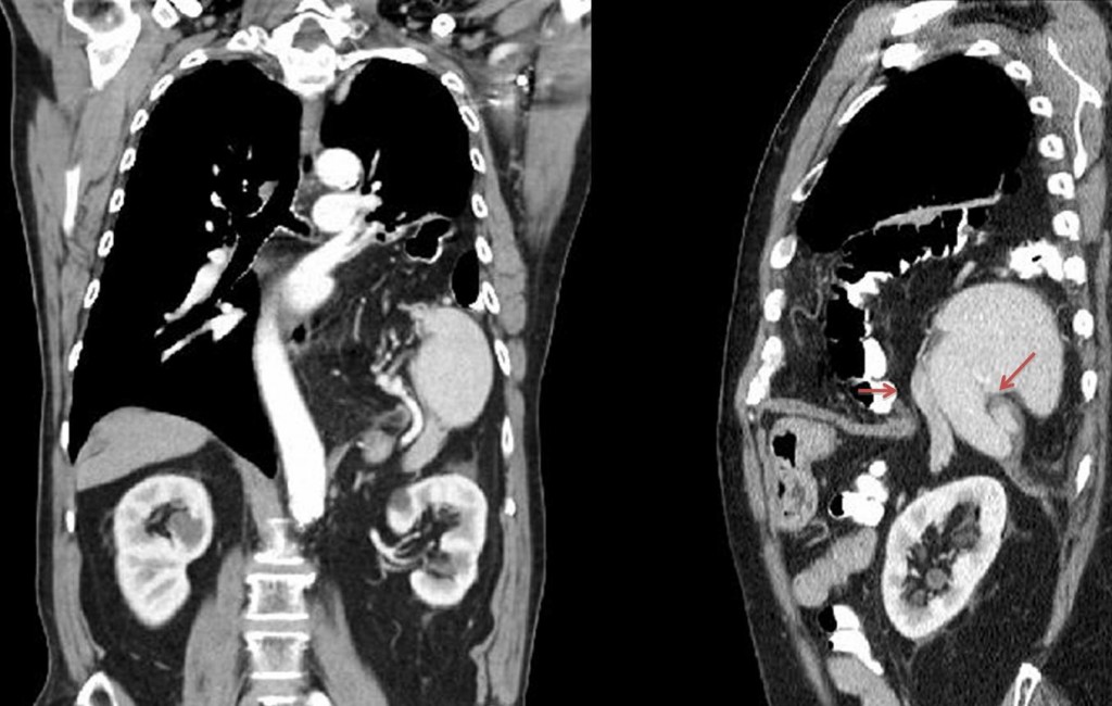
Sagittal and coronal CT show a rent in the diaphragm (arrows) with herniation of the spleen, colon and mesenteric fat.

Final diagnosis: traumatic diaphragmatic hernia.
Muppet is on vacation in Menorca and congratulates the faithful few who participated and answered correctly.
Teaching point: the diaphragm is a frontier organ. Any density in the lower lobes and in contact with the diaphragm may arise from below.


Air densities LLL with no visualization of diaphragm would make intrathoracic herniation of intestinal content my first choice.
Diafragmatic rupture, previous trauma?
Yes
Nel contesto dell’opacamento alla base toracica di sx sono visibili, immagini gassose, sia in AP sia in LL: la DIAGNOSI PIù PROBABILE è ERNIA INTESTINALE DI mORGAGNI-lARREY CON VERSAMENTO PLEURICO REATTIVO. nb: SONO FAN DEL bari (SERIE b ITALIANA) ED ANCH’IO “NORMALE” COME vILLANUEVA!!!!
L’ipotesi di una ernia diaframmatica di morgagni-Larrey è quella che per prima si ipotizza vedendo immagini gassose nel contesto dell’opacamento alla base dell’emitorace sx…. ma non sembra esatta se si conderano due dati: uno clinico(aneurisma aorta addominale) ed uno radiografico( l’aspetto delle raccolte gassose).Si puo’ pertanto ipotizzare un idro-pneumo-mediastino, da “fissuazione” dell’aneurisma e “fistolizzazione” in un’ansa intetstinale, con risalita di contenuto gassoso, attraverso la anatomica continuità tra il retro-peritoneo ed il mediastino.
Elevation of left hemidiaphragma. I would think in multiple situations:
1.post surgical change (I wouldnt expect sucg this elevation and more mediastinal deviation and reactive hiperinsuflation of the rest of lunch parenchyma);
2.atelectasia (i also wouldnt expect this image and moreover I see a “normally located” cisure in the lateral view.
3. Subpulmonary pleural effusion (don’t have to see air images in the fluid interphase)..
4. Diaphragmatic hernia (possible judging the image alone, but I dont find the relation with the aortic aneurism).
5.Diaphragmatic paralysis (I think it could be, then I would look for anterorly located thoracic lesion as possible causes – dont see possible cause in the image but the aortic aneurism depending on its location could cause it by direct compression of the phrenic nerve or even by indirect irritation).
With this I would choose LEFT DIAPHRAGMATIC PARALYSIS (possibly caused by the aortic aneurism).
The story of this patient could be: aortic aneurysm => compression on the hemiazygos vein => chronic pleural effusion only on the left side => repeated drainage = > chronic empyema (air transparency larger on PA-Margulis)
My choice is 3. Chronic empyema
No se visualiza la burbuja gástrica.
Todo sugiere hernia diafragmática postraumatica.
Buscando antiguas fracturas solo se ve raro el esternon en la lateral. El tamaño de la imagen hace dificil valorarla. Muppet podria poner las imagenes del tamaño de sello de correos y aun tendría mas emoción
Muppet is small and like small images. Offers to send you a complimentary magnifying glass.
The left hemidiaphragm is elevated. The trachea is central and there is no mediastinal shift. There are gas-filled bowel loops in the left thorax, suggestive of a left-sided diaphragmatic hernia.
Six answers (five correct), and three lenguages! Muppet is atonished!
Looks like diaphragm rupture. In addition the same in Russian)
Обращает на себя внимание субтотальное затенение легочного поля слева, негомогенное, органы средостения несколько смещены вправо, что может говорить о диафрагмальной грыже или разрыве диафрагмы. Тень левой гемидиафрагмы на этом фоне четко не визуализируется. Если учитывать другие клинические данные, как аневризма абдоминального отдела аорты, то можно предположить травматическое повреждение диафрагмы, а именно ее разрыв.
Quotation: ” The diaphragm is a stupid organ. If something happens to it, it always goes up”.
J.Caceres 😉
order an round baby cribs for sale and check coupon code available bJnZvIdY http://www.best-babycribsforsale.com/