Dear Friends,
Since 100% of you gave correct answers in the previous case, Muppet believes you know it all and is considering retiring to a nunnery. Before he makes his final vows, he wants to show the case of a 35-year-old woman with a solitary calcified lung nodule, discovered in pre-op CT for a gastric tumour.
Diagnosis:
1. Granuloma
2. Chondroma
3. Hamartoma
4. None of the above

35-year-old woman, axial CT
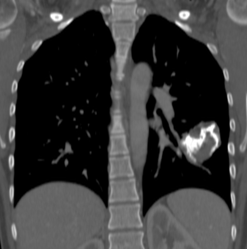
35-year-old woman, coronal CT
Click here for the answer to case 41
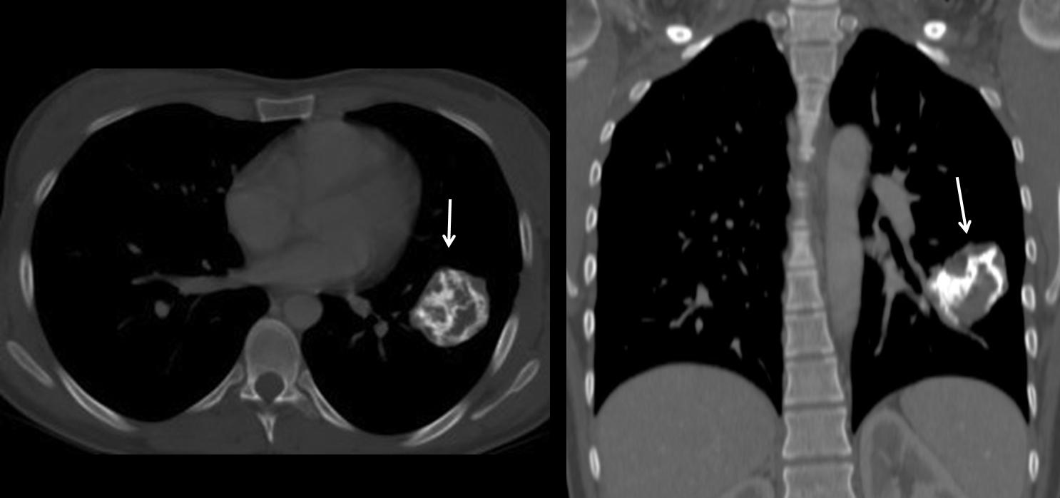
Findings: Axial and coronal CT show a heavily calcified lung nodule (arrows).
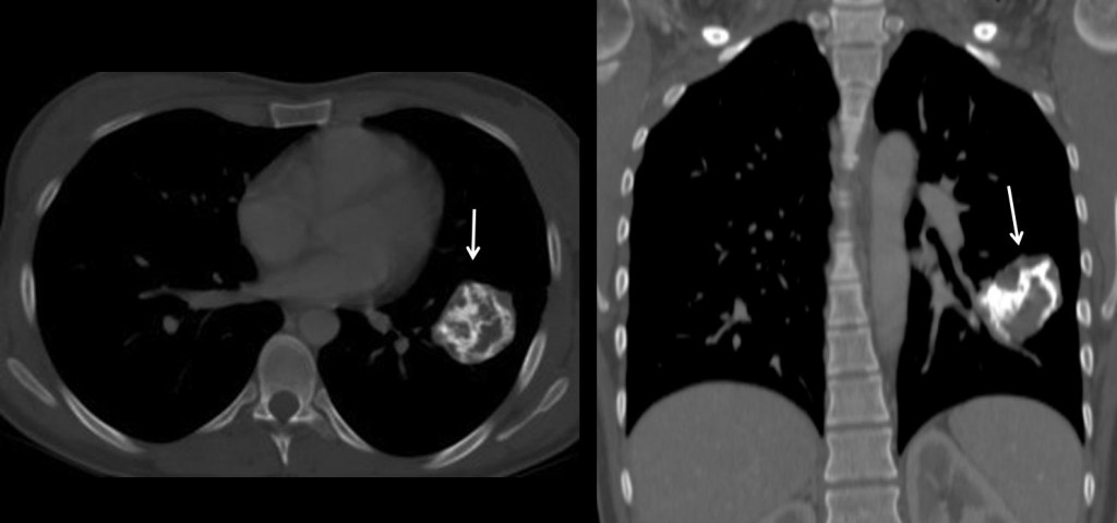
Fig. 1
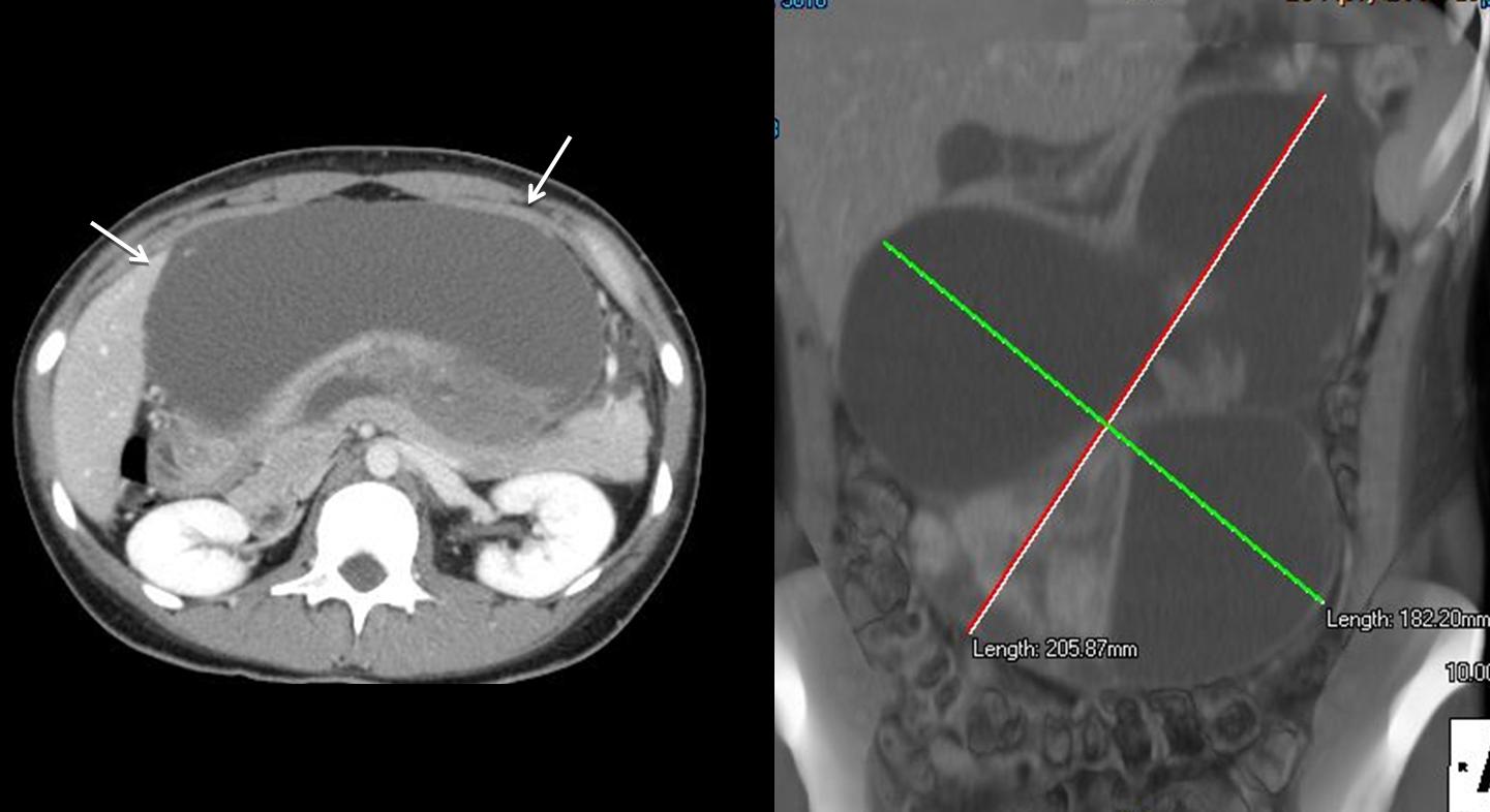
Calcification is lineal and does not look like the amorphous clumps of calcium of hamartoma or granuloma. Abdominal CT shows a large cystic tumour (arrows), proven to be a gastric GIST.
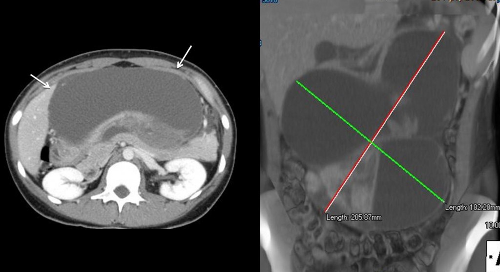
Fig. 2
The combination of such a tumour with a calcified mass in the lung suggests that the patient suffers from a Carney’s triad (GIST, chondroma and paraganglioma). It was elected not to operate on the lung tumour, which, after four years, was unchanged.
Final diagnosis: chondroma of the lung (unproven).
Teaching point: for practical purposes, heavily calcified lung nodules are benign. The differential diagnosis lies between hamartomas and granulomas. Other types, as in the case shown here, are very unusual.