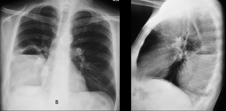
to celebrate the coming year Muppet wants to finish the chapter of air-fluid levels by showing you two patients with fever and malaise.
1. A in a pleural cavity and B in a lung cavity
2. B in a pleural cavity and A in a lung cavity
3. Both in a lung cavity
4. Both in a pleural cavity
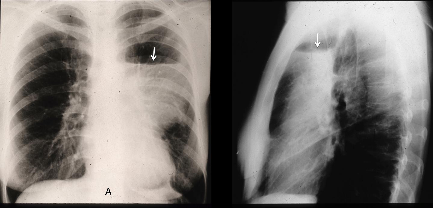
Case A findings: chest films show an air-fluid level (arrows) in the left hemithorax which follows the requisites for hydropneumothorax: the length of air-fluid level is different in PA and lateral projections, has no superior wall and goes across the whole hemithorax in the PA projection.
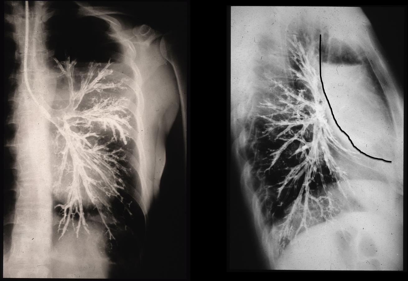
The case is from the pre-CT era and diagnosis was confirmed with broncography, which shows an extrapulmonary lesion displacing the bronchial tree without involvement of bronchi (Muppet performed the study and is very proud of his technique).
Case B shows a right lower peripheral lesion with air-fluid level and a superior wall. At first glance, it seems intrapulmonary. However, there is blunting of the costophrenic angle (arrow) and the lateral view shows a superior obtuse angle, suggestive of an extrapulmonary lesion.
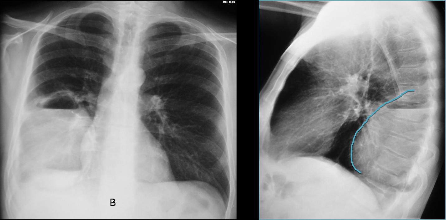
The findings suggest loculated pleural fluid. CT confirms the loculated fluid and the intact lung. Final diagnosis: loculated empyema.
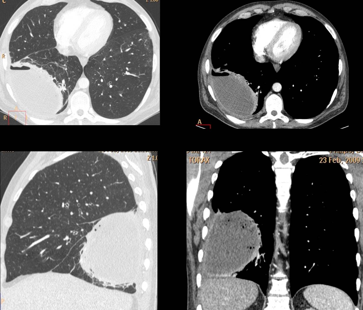
So, the final answer is # 4: both air-fluid levels are in the pleural space
Teaching point: loculated fluid may sometimes simulate a pulmonary cavity because the levels are of equal length and they may have a superior wall. Clues to the correct diagnosis are: peripheral location, blunted costophrenic angle and extrapulmonary contour.