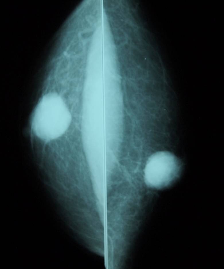
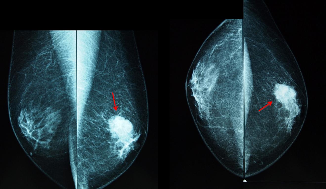
I would like to begin a new subspecialty with the case of a 58-year-old male with a palpable nodule in the left breast.
Findings: Mammograms show a rounded nodule in left breast (arrows), reasonably well defined but with contour irregularities.
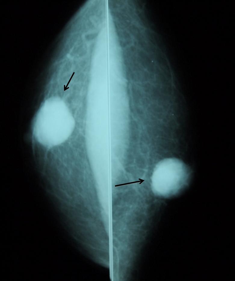
Fig. 2
The most common cause of a palpable lesion in the male breast is gynecomastia. However, in men older than 45 years, the uncommon malignant nodule should always be excluded. When the mammogram shows a nodule, an ultrasound examination should be done. In this particular patient, US confirmed a solid lesion (arrows), and biopsy yielded malignant tissue.
Final diagnosis: intraductal carcinoma of male breast.
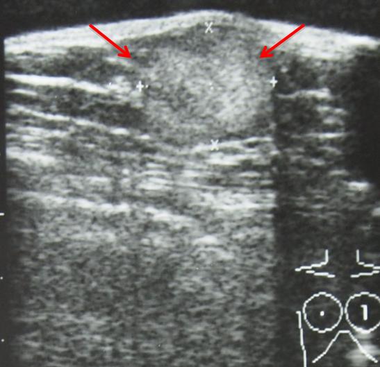
Fig. 3
Carcinoma of the male breast represents less than 1% of all breast neoplasms.
The lesions have the same mammographic and sonographic characteristics as those in females. As occurs in women, the incidence increases with age.
It has been said that their prognosis is poorer than in women, but this is due to the fact that they are often detected at higher stage than women, owing to delay in diagnosis (no screening programs in men).
All histologic types occur; papillary carcinoma is more common in men than in women.
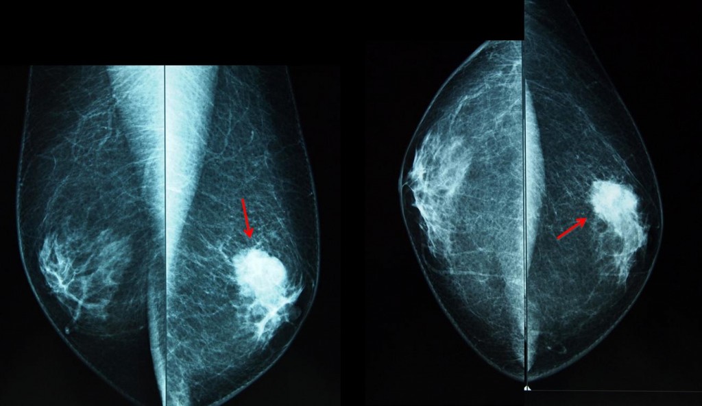
Fig. 4
Fig. 4 is a 53-year-old man with gynecomastia and a palpable left breast nodule (arrows). Note the irregular contours of the nodule, which is located eccentrically from the nipple. Histologic diagnosis: ductal carcinoma.
Differential diagnosis
Gynecomastia is the most common abnormality of the male breast. Nodular gynecomastia should not be confused with a real nodule. Mammography and ultrasound examination will demonstrate the typical appearance of mammary tissue (arrows), excluding further work-up.
There is no evidence that gynecomastia increases the likelihood of malignancy
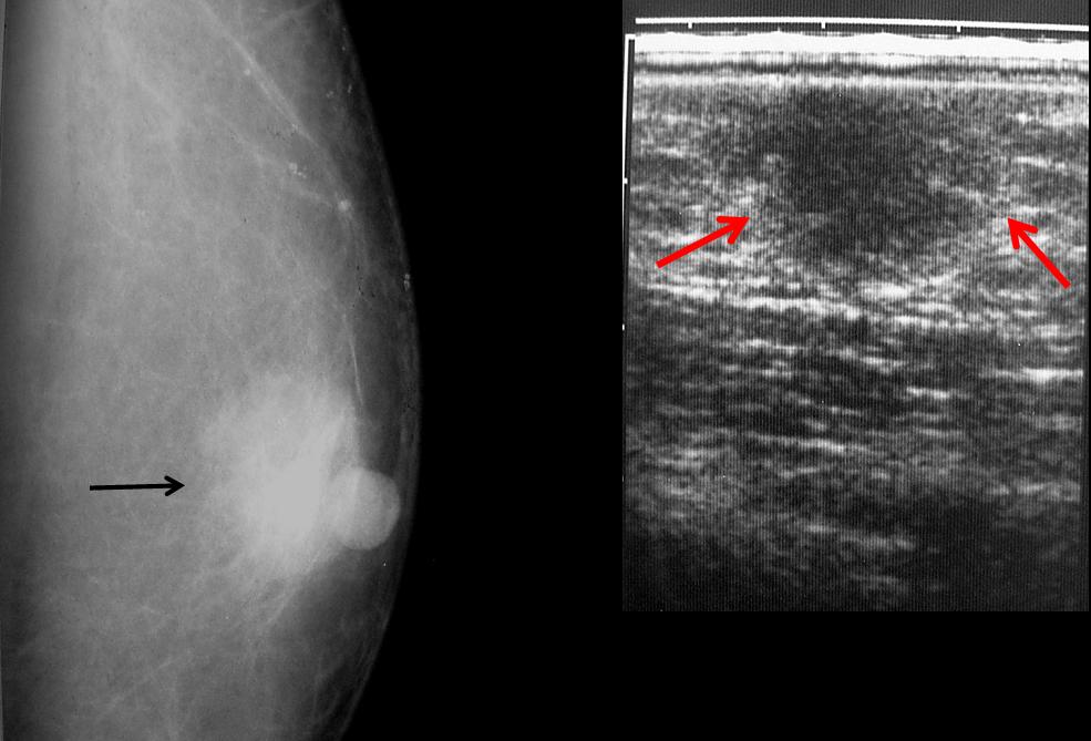
Fig. 5
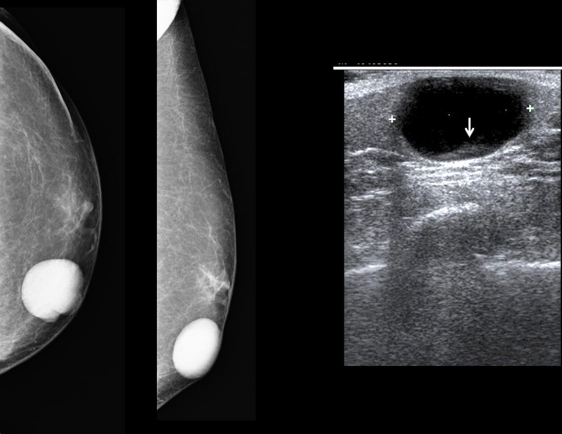
Epidermal inclusion cyst is a dermic tumour that is well-defined and adherent to the skin. Ultrasound confirms the typical cystic appearance, with some debris in the dependent area (arrow).
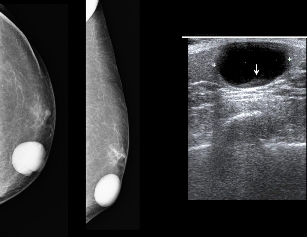
Fig. 6
Fibradenomas are extremely rare in men. The male breast is composed of fat and remnants of ductal tissue; lobular tissue does not develop. Therefore, lesions that originate from the lobules such as fibroadenomas are practically non-existent in the male breast.

Follow Dr. Pepe’s advice
Follow Dr. Pepe’s advice:
Gynecomastia is the most common abnormality of the male breast
A solid breast nodule in a man older than 45 years should be investigated to rule out malignancy
Imaging manifestations of breast carcinoma in men are the same as in women
Suggested reading: Imaging characteristics of malignant lesions of the male breast. RadioGraphics 26:993-1006, 2006
Case prepared by Elena Rabanal, MD