Dr. Pepe’s Diploma Casebook: Case 17 – SOLVED!
Dear friends,
to welcome the new year I am showing you the radiographs of a 70 year-old male being evaluated for possible lung metastases.
Diagnosis:
1. Calcified pericardium
2. Ventricular aneurysm
3. Cardiac myxoma
4. None of the above
Findings:
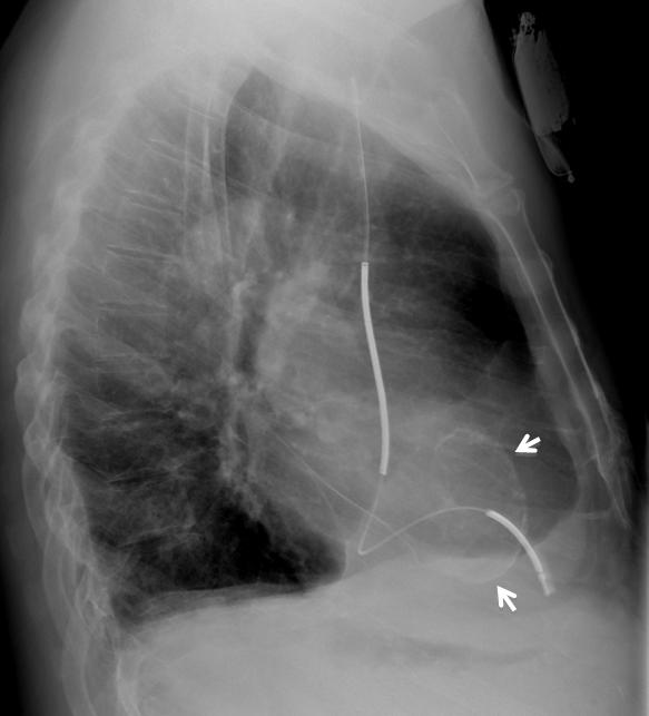
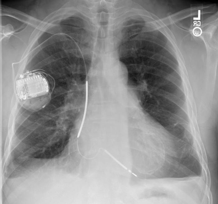
There are no metastases. The heart is enlarged in a left ventricular configuration but the vessels are not distended. There is a circular calcification overlying the cardiac silhouette (arrows) and scarring in the costophrenic angles.

Fig. 1
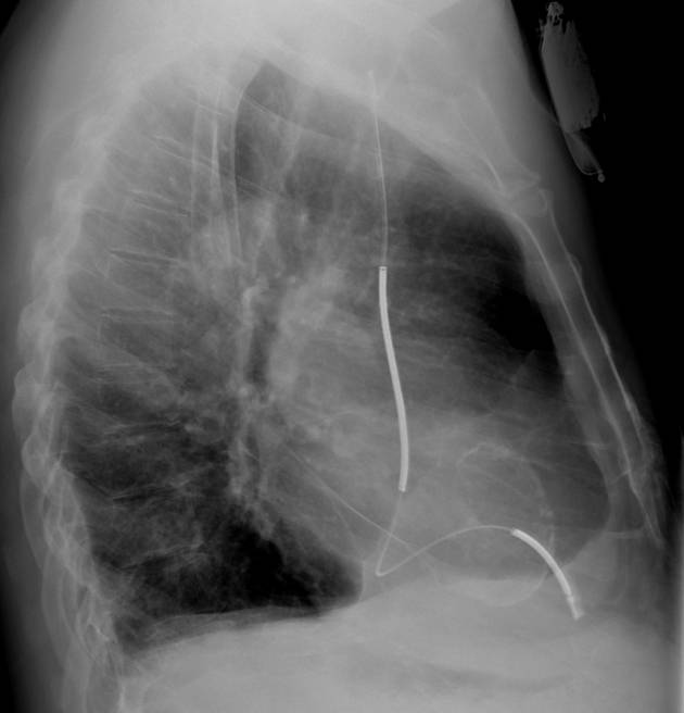
In the lateral view, the circular calcification projects within the cardiac contour (arrows).

Fig. 2
Axial CT confirms the location within the left ventricle (arrow). The low density area deep to the calcification (*) indicates longstanding organized thrombus.

Fig. 3
Diagnosis: Calcified left ventricular aneurysm
Discussion:
Left ventricular aneurysm usually follows myocardial infarction. Initially, the affected portion of myocardium is dyskinetic or akinetic; subsequently, there is often progression to paradoxical motion (bulging out when the remaining myocardium contracts in systole). This condition often leads to sustained left heart failure. Our patient was not in failure at this time. On CT, the damaged muscle is often thin or may bulge in systole. Calcification indicates a remote infarct. Left ventricular clot may form and either organize or embolize. An MRI (Fig. 4) on a different patient shows an apical aneurysm. Note apical bulge, thinning of muscle (arrow), and absence of clot.

Fig. 4
The initial diagnosis is usually made with x-ray, CT, or echocardiography. The physiologic function of the left ventricle can be evaluated with several modalities, including multi-slice CT, echocardiography, ventricular scintigraphy, and MRI. The degree of calcification and the degree of muscle damage are not necessarily the same.
Left ventricular pseudo (false) aneurysms are usually found in a posterior and inferior location, whereas true aneurysms tend to be anterior and lateral. Pseudoaneurysms tend to rupture and, therefore, require urgent surgical resection, whereas true aneurysms rarely rupture.
Differential Diagnosis:
Pericardial calcification (Fig. 5 & 6) can usually be differentiated from myocardial calcification. It is visible on the cardiac surface in some projections and more often affects the right and anterior cardiac silhouette. Calcification does not necessarily lead to constrictive pericarditis.

Fig. 5
Coarse pericardial calcification in PA and lateral views (Fig.5 A,B arrows). Coronal and axial CT confirm the peripheral location of the calcium (Fig. 6 A,B arrows).

Fig. 6

Follow Dr. Pepe’s advice
Follow Dr. Pepe’s advice:
- Calcified ventricular aneurysm presents as a thin curvilinear calcification located at the tip of the heart. Early on, there may just be thinning of the myocardium. Also look for thrombus.
- True aneurysms are anterior and seldom rupture , while false aneurysms are posterior, tend not to calcify and do ruputre.
- Pericardial calcification is coarse and usually affects the right side of the cardiac border.
Suggested reading: “Evaluation of Global and Regional Left Ventricular Function With 16-Slice Computed Tomography, Biplane Cineventriculography, and Two-Dimensional Transthoracic Echocardiography: Comparison With Magnetic Resonance Imaging.” J Am Coll Cardiol 48(10): 2034-2044.
Case prepared by Lawrence Goodman MD


La calficazione è a “guscio d’uovo” cioè periferica e non dimostra eventuale “core” centrale come da una massa: essa “disegna” la valvola mitralica, calcificata in toto, come nella BIG-MAC ( Massiva Mitralica Annulus Calcification).Essa compare nel 6/° delle persone sopra i 60 anni.Una sua variante è la Necrosi Caseosa.La diagnosi, sospettata sull’Rx-torace, può essere confermata da una ecocardiografia e/o RM.
– oval lesion in the left ventricle with calcified walls (calcified parietal cyst, echinococcosis?)
– bilateral pleural effusion
– pacemaker
Ventricular aneurysm
Calcified left ventricular aneurysm (post-infarction). They do not necessarily “bulge”.
Mild pulmonary hypertension.
Residual bilateral pleural thickening.
mitral annulus calcification
– Pericardial calcification could be related to hydatid cyst or TB infection.
Will you define the calcification as thin or thick-walled?
It’s quite thin…almost eggshell like.
I was thinking along the lines of a calcified left ventricular aneurysm.
To me it does not look like calcification of the mitral annulus.
Nor it does to me. Answer next week. Happy new year!
Ventricular aneurysm
ventricular aneurysm